Updated on 2024/04/24

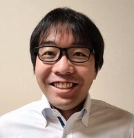
写真a

 
MORIMOTO, Yuya
 
Affiliation
Faculty of Science and Engineering, School of Fundamental Science and Engineering
Job title
Associate Professor
Degree
博士(情報理工学) ( 東京大学 )

Research Experience

  • 2023.04
    -
    Now

    Waseda University   Faculty of Science and Engineering   Associate Professor

  • 2019.04
    -
    2023.03

    The University of Tokyo

  • 2014.04
    -
    2019.03

    The University of Tokyo   Institute of Industrial Science

  • 2014.06
    -
    2016.03

    科学技術振興機構   ERATO竹内バイオ融合プロジェクト   研究総括補佐・グループリーダー

  • 2009.04
    -
    2011.03

    富士フイルム株式会社   メディカルシステム開発センター

Education Background

  • 2011.04
    -
    2014.03

    東京大学大学院   情報理工学系研究科   知能機械情報学専攻 博士課程  

  • 2007.04
    -
    2009.03

    東京大学大学院   情報理工学系研究科   知能機械情報学専攻 修士課程  

Committee Memberships

  • 2023.04
    -
    Now

    日本機械学会 マイクロナノ工学部門  部門幹事

  • 2021.10
    -
    Now

    日本機械学会 マイクロナノ工学部門 未来のセンサに関する研究会  幹事

  • 2021.10
    -
    Now

    電気学会 E部門BMS技術委員会 未来のセンサ調査専門委員会  幹事補佐

  • 2020.04
    -
    Now

    電気学会 センサ・マイクロマシン部門大会 「センサ・マイクロマシンと応用システムシンポジウム」  論文委員

  • 2020.04
    -
    Now

    電気学会 デジタル・バイオ融合調査専門委員会  委員

  • 2020.04
    -
    Now

    電気学会 立体構造微細加工技術と異分野融合によるライフサイエンス応用と実用化に関する調査専門委員会  委員

  • 2017.04
    -
    Now

    日本機械学会 マイクロ・ナノ工学部門 マイクロ・ナノ工学シンポジウム  実行委員

  • 2022.04
    -
    2023.03

    日本機械学会 マイクロナノ工学部門 広報委員会  委員長

  • 2021.04
    -
    2023.03

    日本機械学会 マイクロナノ工学部門 運営委員会  委員

  • 2021.04
    -
    2022.03

    日本機械学会 マイクロナノ工学部門 広報委員会  幹事

  • 2017.07
    -
    2019.08

    日本機械学会 マイクロ・ナノ工学部門 マイクロ・ナノ医療デバイスに関する研究会  幹事

  • 2017.07
    -
    2019.08

    電気学会 E部門 BMS技術委員会 マイクロ・ナノバイオ医療デバイス調査専門委員会  幹事

  • 2015.04
    -
    2017.03

    電気学会 E部門 BMS技術委員会 材料から革新するバイオマイクロシステム調査専門委員会  委員

  • 2016.06


    Science Outreach Event, the 20th MicroTAS conference  Assistant

  • 2015.10
    -
    2016.04

    化学とマイクロ・ナノシステム学会 第33回研究会  実行委員

▼display all

Research Areas

  • Nano/micro-systems / Mechanics and mechatronics / Robotics and intelligent system

Research Interests

  • マイクロ流体

  • マイクロ・ナノデバイス

  • 再生医療

  • 組織工学

Awards

  • 船井情報科学振興財団 船井学術賞

    2022.05  

  • 日本機械学会 日本機械学会論文賞

    2022.04  

  • 令和3年度文部科学大臣表彰 若手科学者賞

    2021.04  

  • Young Researcher Poster Award

    2020.10   The 24th International Conference on Miniaturized Systems for Chemistry and Life Science (MicroTAS2020)  

    Winner: Byeongwook Cho, Yuya Morimoto, Shoji Takeuchi

  • 新分野開拓表彰

    2020.10   日本機械学会マイクロ・ナノ工学部門  

    Winner: 森本雄矢

  • 8th Advanced Robotics Paper Awards

    2020.10   Advanced Robotics Best Paper Awards

  • 平成30年度日本機械学会奨励賞(研究)

    2019.04   日本機械学会  

    Winner: 森本雄矢

  • 平成29年度若手優秀賞

    2018.03   化学とマイクロ・ナノシステム学会  

    Winner: 森本雄矢

  • Outstanding Student Paper Award Finalist

    2017.01   The 30th IEEE International Conference on Micro Electro Mechanical Systems (MEMS2017)  

    Winner: Yusuke Hirata, Yuya Morimoto, Eunyel Nam, Shotaro Yoshida, Shoji Takeuchi

  • 弥生賞

    2014.07   東京大学生産技術研究所  

    Winner: 森本雄矢

  • 研究科長賞(博士)

    2014.03   東京大学大学院 情報理工学系研究科  

    Winner: 森本雄矢

  • 総長賞

    2014.03   東京大学  

    Winner: 森本雄矢

  • 平成25年優秀論文発表賞A

    2014.02   電気学会  

    Winner: 森本雄矢

  • 第4回育志賞

    2014.02   日本学術振興会  

    Winner: 森本雄矢

  • 奨励賞

    2013.11   電気学会 E部門 第30回「センサ・マイクロマシンと応用システム」シンポジウム  

    Winner: 森本雄矢

  • Young Researcher Poster Award

    2013.10   The 17th International Conference on Miniaturized Systems for Chemistry and Life Science (MicroTAS2013)  

    Winner: 森本雄矢

  • Outstanding Oral Paper Award

    2013.01   The 26th IEEE International Conference on Micro Electro Mechanical Systems (MEMS2013)  

    Winner: 森本雄矢

  • ポスター賞

    2012.06   第12回東京大学生命科学シンポジウム  

    Winner: 森本雄矢

  • 研究科長賞 (修士)

    2009.03   東京大学大学院 情報理工学系研究科  

    Winner: 森本雄矢

▼display all


Papers

  • Biohybrid tensegrity actuator driven by selective contractions of multiple skeletal muscle tissues

    Kazuma Morita, Yuya Morimoto, Shoji Takeuchi

    Biofabrication   15 ( 4 )  2023.10

     View Summary

    Biohybrid robots are robots composed of both biological and artificial materials that can exhibit the unique characteristics commonly found in living organisms. Skeletal muscle tissues can be utilized as their actuators due to their flexibility and ON/OFF controllability, but previous muscle-driven robots have been limited to one-degree of freedom (DOF) or planar motions due to their design. To overcome this limitation, we propose a biohybrid actuator with a tensegrity structure that enables multiple muscle tissues to be arranged in a 3D configuration with balanced tension. By using muscle tissues as tension members of tensegrity structure, the contraction of muscle tissues can cause the movement of the actuator in multiple-DOFs. We demonstrate the fabrication of the biohybrid tensegrity actuator by attaching three cultured skeletal muscle tissue made from C2C12 cells and fibrin-based hydrogel to an actuator skeleton using a snap-fit mechanism. When we applied an electric field of more than 4 V mm−1 to the skeletal muscle tissue, the fabricated actuator had a structure to tilt in multiple directions through the selective displacement of about 0.5 mm in a specific direction caused by the contractions of muscle tissue, resulting in 3D multi-DOF tilting motion. We also show that the actuator possesses superior characteristics of tensegrity structure such as stability and robustness by assessing the response of the actuator to external force. This biohybrid tensegrity actuator provides a useful platform for the development of muscle-driven biohybrid robots with complex and flexible movements.

    DOI PubMed

    Scopus

    2
    Citation
    (Scopus)
  • Real-time quantitative characterization of ion channel activities for automated control of a lipid bilayer system.

    Kazuto Ogishi, Toshihisa Osaki, Hisatoshi Mimura, Izumi Hashimoto, Yuya Morimoto, Norihisa Miki, Shoji Takeuchi

    Biosensors & bioelectronics   237   115490 - 115490  2023.06  [International journal]

     View Summary

    This paper describes a novel signal processing method to characterize the activity of ion channels on a lipid bilayer system in a real-time and quantitative manner. Lipid bilayer systems, which enable single-channel level recordings of ion channel activities against physiological stimuli in vitro, are gaining attention in various research fields. However, the characterization of ion channel activities has heavily relied on time-consuming analyses after recording, and the inability to return the quantitative results in real time has long been a bottleneck to incorporating the system into practical products. Herein, we report a lipid bilayer system that integrates real-time characterization of ion channel activities and real-time response based on the characterization result. Unlike conventional batch processing, an ion channel signal is divided into short segments and processed during the recording. After optimizing the system to maintain the same characterization accuracy as conventional operation, we demonstrated the usability of the system with two applications. One is quantitative control of a robot based on ion channel signals. The velocity of the robot was controlled every second, which was around tens of times faster than the conventional operation, in proportion to the stimulus intensity estimated from changes in ion channel activities. The other is the automation of data collection and characterization of ion channels. By constantly monitoring and maintaining the functionality of a lipid bilayer, our system enabled continuous recording of ion channels over 2 h without human intervention, and the time of manual labor has been reduced from conventional 3 h to 1 min at a minimum. We believe the accelerated characterization and response in the lipid bilayer systems presented in this work will facilitate the transformation of lipid bilayer technology toward a practical level, finally leading to its industrialization.

    DOI PubMed

    Scopus

    1
    Citation
    (Scopus)
  • Co-culture system of human skin equivalents with mouse neural spheroids

    Satoshi Inagaki, Yuya Morimoto, Ikuo K. Suzuki, Kazuo Emoto, Shoji Takeuchi

    Journal of Bioscience and Bioengineering    2023

     View Summary

    This study describes a co-culture system of human skin equivalents (HSEs) and dorsal root ganglion (DRG) neurons. We prepared spheroids of mouse DRG neurons with or without Schwann cells (SCs). Spheroids comprising DRG neurons and SCs showed longer neurite extensions than those comprising DRG neurons alone. Neurite extension of more than 1 mm was observed from spheroids cultured inside HSEs, whereas neurite extension was primarily observed on the surface of HSEs from spheroids cultured on HSEs. We propose that our model may be a useful tool for studying neurite extension in the human skin.

    DOI PubMed

    Scopus

  • Microfluidic Device to Manipulate 3D Human Epithelial Cell-Derived Intestinal Organoids

    Miki Matsumoto, Yuya Morimoto, Toshiro Sato, Shoji Takeuchi

    Micromachines   13 ( 12 )  2022.12

     View Summary

    In this study, we propose a microfluidic organoid-trapping device used to immobilize human intestinal organoids and apply fluidic stimuli to them. The proposed device has a microchannel with a trapping region with wall gaps between the channel walls and the bottom surface, and a constriction to clog the organoids in the channel. Since the introduced culture medium escapes from the gap, organoids can be cultured without excessive deformation by hydrostatic pressure. Owing to the characteristics of the organoid-trapping device, we succeeded in trapping human intestinal organoids in the channel. Furthermore, to demonstrate the applicability of the device for culturing intestinal organoids, we induced organoid fusion to form large organoids by aligning the organoids in the channel and applying fluidic shear stress to the organoids to regulate their surface structures. Therefore, we believe that organoid-trapping devices will be useful for investigating organoids aligned or loaded with fluidic stimulation.

    DOI

    Scopus

  • Living skin on a robot

    Michio Kawai, Minghao Nie, Haruka Oda, Yuya Morimoto, Shoji Takeuchi

    Matter   5 ( 7 ) 2190 - 2208  2022.07

     View Summary

    Humanoids are robots created with human forms or characteristics; these robots also have the potential to seamlessly interact with human beings. By replicating the appearances and functions (e.g., self-healing) of human beings, humanoids have the potential to establish more harmonic and natural human-robot interactions. Here, we propose the use of skin equivalent, a living skin model consisting of cells and extracellular matrix, as a human-like and self-healing coverage material for robots. We fabricated a three-joint robotic finger covered with skin equivalent by developing a method to cover three-dimensional objects with skin equivalent. Furthermore, inspired by the medical treatment of deeply burned skin using grafted hydrogels, we demonstrated wound repair of a dermis equivalent covering a robotic finger by culturing the wounded tissue grafted with a collagen sheet. With the above results, this research shows the potential of using skin equivalent as human-like and self-healing coverage material for robots.

    DOI

    Scopus

    14
    Citation
    (Scopus)
  • 3D printed microfluidic devices for lipid bilayer recordings

    Kazuto Ogishi, Toshihisa Osaki, Yuya Morimoto, Shoji Takeuchi

    Lab on a Chip   22 ( 5 ) 890 - 898  2022.03

     View Summary

    This paper verifies the single-step and monolithic fabrication of 3D structural lipid bilayer devices using stereolithography. Lipid bilayer devices are utilized to host membrane proteins in vitro for biological assays or sensing applications. There is a growing demand to fabricate functional lipid bilayer devices with a short lead-time, and the monolithic fabrication of components by 3D printing is highly anticipated. However, the prerequisites of 3D printing materials which lead to reproducible lipid bilayer formation are still unknown. Here, we examined the feasibility of membrane protein measurement using lipid bilayer devices fabricated by stereolithography. The 3D printing materials were characterized and the surface smoothness and hydrophobicity were found to be the relevant factors for successful lipid bilayer formation. The devices were comparable to the ones fabricated by conventional procedures in terms of measurement performances like the amplitude of noise and the waiting time for lipid bilayer formation. We further demonstrated the extendibility of the technology for the functionalization of devices, such as incorporating microfluidic channels for solution exchangeability and arraying multiple chambers for robust measurement. This journal is

    DOI PubMed

    Scopus

    12
    Citation
    (Scopus)
  • Functional analysis of human brain endothelium using a microfluidic device integrating a cell culture insert.

    Shigenori Miura, Yuya Morimoto, Tomomi Furihata, Shoji Takeuchi

    APL bioengineering   6 ( 1 ) 016103 - 016103  2022.03  [International journal]

     View Summary

    The blood-brain barrier (BBB) is a specialized brain endothelial barrier structure that regulates the highly selective transport of molecules under continuous blood flow. Recently, various types of BBB-on-chip models have been developed to mimic the microenvironmental cues that regulate the human BBB drug transport. However, technical difficulties in complex microfluidic systems limit their accessibility. Here, we propose a simple and easy-to-handle microfluidic device integrated with a cell culture insert to investigate the functional regulation of the human BBB endothelium in response to fluid shear stress (FSS). Using currently established immortalized human brain microvascular endothelial cells (HBMEC/ci18), we formed a BBB endothelial barrier without the substantial loss of barrier tightness under the relatively low range of FSS (0.1-1 dyn/cm2). Expression levels of key BBB transporters and receptors in the HBMEC/ci18 cells were dynamically changed in response to the FSS, and the effect of FSS reached a plateau around 1 dyn/cm2. Similar responses were observed in the primary HBMECs. Taking advantage of the detachable cell culture insert from the device, the drug efflux activity of P-glycoprotein (P-gp) was analyzed by the bidirectional permeability assay after the perfusion culture of cells. The data revealed that the FSS-stimulated BBB endothelium exhibited the 1.9-fold higher P-gp activity than that of the static culture control. Our microfluidic system coupling with the transwell model provides a functional human BBB endothelium with secured transporter activity, which is useful to investigate the bidirectional transport of drugs and its regulation by FSS.

    DOI PubMed

    Scopus

    6
    Citation
    (Scopus)
  • Skeletal muscle-adipose cocultured tissue fabricated using cell-laden microfibers and a hydrogel sheet

    Byeongwook Jo, Yuya Morimoto, Shoji Takeuchi

    Biotechnology and Bioengineering   119 ( 2 ) 636 - 643  2022.02

     View Summary

    The emerging interest in skeletal muscle tissue originates from its unique properties that control body movements. In particular, recent research advances in engineered skeletal muscle tissue have broadened the possibilities of applications in nonclinical models. However, due to the lack of adipose tissue, current engineered skeletal muscle tissue has the limitation of satisfying in vivo-like position and proportion of intermuscular fat. Adipose tissue within the skeletal muscle affects their functional properties. Here, a fabrication method for cocultured tissue composed of skeletal muscle and adipose tissues is proposed to reproduce the functional and morphological characteristics of muscle. By implementing prematured adipose microfibers in a myoblast-laden hydrogel sheet, both the accumulation of large lipid droplets and control of the position of adipose tissue within the skeletal muscle tissue becomes feasible. The findings of this study provide helpful information regarding engineered skeletal muscle, which has strong potential in drug screening models.

    DOI PubMed

    Scopus

    6
    Citation
    (Scopus)
  • Biohybrid Soft Robots Driven by Contractions of Skeletal Muscle Tissue

    Yuya Morimoto, Shoji Takeuchi

    Journal of Robotics and Mechatronics   34 ( 2 ) 260 - 262  2022  [Refereed]

     View Summary

    In this letter, we introduce biohybrid robots powered by skeletal muscle tissue. Culturing myoblast-laden extracellular matrix structures enables the construc-tion of skeletal muscle tissue in vitro. Biohybrid robots constructed by the integration of such fabricated muscle tissue with robot skeletons have achieved vari-ous movements, according to the configuration of the skeleton. We believe that biohybrid robots will in-creasingly become available in the field of robotics.

    DOI

    Scopus

    2
    Citation
    (Scopus)
  • 3D-Printed Centrifugal Pump Driven by Magnetic Force in Applications for Microfluidics in Biological Analysis

    Byeongwook Jo, Yuya Morimoto, Shoji Takeuchi

    Advanced Healthcare Materials   11 ( 24 )  2022

     View Summary

    In recent years, microfluidic systems have been extensively utilized for biological analysis. The integration of pumps in microfluidic systems requires precise control of liquids and effort-intensive set-ups for multiplexed experiments. In this study, a 3D-printed centrifugal pump driven by magnetic force is presented for microfluidics and biological analysis. The permanent magnets implemented in the centrifugal pump synchronized the rotation of the driving and operating parts. Precise control of the flow rate and a wide range and variety of flow profiles are achieved by controlling the rotational speed of the motor in the driving part. The compact size and contactless driving part allow simple set-ups within commercially available culture dishes and tubes. It is demonstrated that the fabricated 3D-printed centrifugal pump can induce laminar flow in a microfluidic device, perfusion culture of in vitro tissues, and alignment of cells under shear stress. This device has a high potential for applications in microfluidic devices and perfusion culture of cells.

    DOI PubMed

    Scopus

    4
    Citation
    (Scopus)
  • Microfluidic system for applying shear flow to endothelial cells on culture insert with collagen vitrigel membrane

    Yuya Morimoto, Shogo Nagata, Miki Matsumoto, Keisuke Sugahara, Shigenori Miura, Shoji Takeuchi

    Sensors and Actuators B: Chemical   348   130675 - 130675  2021.12

    DOI

    Scopus

    7
    Citation
    (Scopus)
  • Formation of contractile 3D bovine muscle tissue for construction of millimetre-thick cultured steak

    Mai Furuhashi, Yuya Morimoto, Ai Shima, Futoshi Nakamura, Hiroshi Ishikawa, Shoji Takeuchi

    npj Science of Food   5 ( 1 )  2021.12  [Refereed]

     View Summary

    Owing to the increase in the global demand of meat, cultured meat technology is being developed to circumvent a shortage of meat in the future. However, methods for construction of millimetre-thick bovine muscle tissues with highly aligned myotubes have not yet been established. Here, we propose a culture method for constructing 3D-cultured bovine muscle tissue containing myotubes aligned along its long-axial direction, which contracted in response to electrical stimulation. First, we optimised the composition of biomaterials used in the construction and the electrical stimulation applied to the tissue during culture. Subsequently, we fabricated millimetre-thick bovine muscle tissues containing highly aligned myotubes by accumulating bovine myoblast-laden hydrogel modules. The microbial content of the bovine muscle tissue cultured for 14 days was below the detection limit, indicating that the muscle tissues were sterile, unlike commercial meat. Therefore, the proposed construction method for bovine muscle tissues will be useful for the production of clean cultured steak meat simulating real meat.

    DOI

    Scopus

    79
    Citation
    (Scopus)
  • A Cylindrical Molding Method for the Biofabrication of Plane-Shaped Skeletal Muscle Tissue

    Minghao Nie, Ai Shima, Kenta Fukushima, Yuya Morimoto, Shoji Takeuchi

    Micromachines   12 ( 11 ) 1411 - 1411  2021.11

     View Summary

    Muscle tissues can be fabricated in vitro by culturing myoblast-populated hydrogels. To counter the shrinkage of the myoblast-populated hydrogels during culture, a pair of anchors are generally utilized to fix the two ends of the hydrogel. Here, we propose an alternative method to counter the shrinkage of the hydrogel and fabricate plane-shaped skeletal muscle tissues. The method forms myoblast-populated hydrogel in a cylindrical cavity with a central pillar, which can prevent tissue shrinkage along the circumferential direction. By eliminating the usages of the anchor pairs, our proposed method can produce plane-shaped skeletal muscle tissues with uniform width and thickness. In experiments, we demonstrate the fabrication of plane-shaped (length: ca. 10 mm, width: 5~15 mm) skeletal muscle tissue with submillimeter thickness. The tissues have uniform shapes and are populated with differentiated muscle cells stained positive for myogenic differentiation markers (i.e., myosin heavy chains). In addition, we show the assembly of subcentimeter-order tissue blocks by stacking the plane-shaped skeletal muscle tissues. The proposed method can be further optimized and scaled up to produce cultured animal products such as cultured meat.

    DOI

    Scopus

    6
    Citation
    (Scopus)
  • Living Skin as a Self-Repairable Covering Material for Robots

    Michio Kawai, Minghao Nie, Haruka Oda, Yuya Morimoto, Shoji Takeuchi

    2021 IEEE 34th International Conference on Micro Electro Mechanical Systems (MEMS)    2021.01

    DOI

  • Cell-Based Biohybrid Sensor Device for Chemical Source Direction Estimation

    H. Oda, K. Kihara, Y. Morimoto, S. Takeuchi

    Cyborg and Bionic Systems    2021.01

    DOI

    Scopus

    14
    Citation
    (Scopus)
  • Biohybrid robot with skeletal muscle tissue covered with a collagen structure for moving in air

    Yuya Morimoto, Hiroaki Onoe, Shoji Takeuchi

    APL Bioengineering   4 ( 2 ) 026101 - 026101  2020.06  [Refereed]

    DOI

  • A dynamic microarray device for pairing and electrofusion of giant unilamellar vesicles

    Keisuke Sugahara, Yuya Morimoto, Sho Takamori, Shoji Takeuchi

    Sensors and Actuators, B: Chemical   311  2020.05  [Refereed]

     View Summary

    In this paper, we present a microfluidic device that enables the efficient pairing of giant unilamellar vesicles (GUVs) and electrofusion of paired GUVs. The device consists of a hydrodynamic microarray system for the deterministic pairing of GUVs and two thick microelectrodes, made with a low melting-point fusible alloy, for applying the electric field on the paired GUVs. We achieved 75 % efficiency for pairing GUVs with the dynamic microarray device, which was designed by computing fluid resistance of the channels. We successfully performed electrofusion of the paired GUVs arrayed in the pairing channels. Furthermore, we confirmed that the lipid component and the inner solution of the paired GUVs were fused by electrofusion respectively. We believe our microfluidic device will become a useful tool for studying biological reactions in membrane compartments.

    DOI

    Scopus

    8
    Citation
    (Scopus)
  • Centrifuge-based step emulsification device for simple and fast generation of monodisperse picoliter droplets

    Dong Chel Shin, Yuya Morimoto, Jun Sawayama, Shigenori Miura, Shoji Takeuchi

    Sensors and Actuators, B: Chemical   301  2019.12  [Refereed]

     View Summary

    © 2019 Monodisperse picoliter droplets have been widely used for biochemical reactors and preparation of functional microbeads for food, cosmetics, and medical industry. Although conventional microfluidic technologies enable the production of monodisperse picoliter droplets, special instruments (e.g. syringe pump) are required, leading to its poor usability and accessibility in other research fields. In this study, we propose centrifuge-based step emulsification device housed in a microtube for simple and fast generation of monodisperse picoliter droplets. Our device consists of a reservoir part for storage of the dispersed phase and a microchannel part for droplet formation. Centrifugation using a commercial centrifuge exerts excessive pressure on the dispersed phase, infusing it into a microchannel. The infused dispersed phase is pinched off, forming droplets due to the channel geometry for step emulsification. The produced droplets at the step then are transported to the bottom of the microtube due to the centrifugal force. Using this device, we achieved formation of monodisperse water-in-oil (W/O) picoliter droplets with diameters ranging from 18 to 90 μm. Moreover, we demonstrated the cell-free protein synthesis reaction in monodisperse picoliter droplets as well as the production of cell-sized glucose-responsive hydrogel microbeads. We believe that our device would be a powerful tool for simple and fast preparation of picoliter samples with high usability.

    DOI

    Scopus

    21
    Citation
    (Scopus)
  • 1
    Citation
    (Scopus)
  • Portable biohybrid odorant sensors using cell-laden collagen micropillars.

    Yusuke Hirata, Yuya Morimoto, Eunryel Nam, Shoji Takeuchi

    Lab on a chip   19 ( 11 ) 1971 - 1976  2019.06  [International journal]

     View Summary

    Biohybrid odorant sensors (BOSs) composed of biological materials and artificial detectors have recently attracted much attention due to their high degree of sensitivity and selectivity. Although portability is crucial for the practical use of BOSs on site, the currently used artificial detectors for biological signals are unportable. In this study, we propose a portable cell-based odorant sensor, which uses cell-laden collagen micropillars to compensate the low optical abilities of portable artificial detectors. The micropillars were composed of HEK293T cells expressing olfactory receptors, which emit a fluorescence signal based on the extent of odorant stimulation using a calcium fluorescent indicator. By stacking cells vertically in the micropillars, we achieved different levels of amplification of the fluorescence signals by varying the height of the micropillars. As a working demonstration of the portable BOS, we successfully detected different concentrations of odorants using an inexpensive web camera. The BOS was also able to distinguish the slight differences between an agonist and an antagonist. We believe that the portability of our BOS would facilitate its applications in point-of-care testing and on-site detection of hazardous materials.

    DOI PubMed

    Scopus

    13
    Citation
    (Scopus)
  • Temporal Observation of Adipocyte Microfiber Using Anchoring Device.

    Akiyo Yokomizo, Yuya Morimoto, Keigo Nishimura, Shoji Takeuchi

    Micromachines   10 ( 6 )  2019.05  [International journal]

     View Summary

    In this paper, we propose an anchoring device with pillars to immobilize an adipocyte microfiber that has a fiber-shaped adipocyte tissue covered by an alginate gel shell. Because the device enabled the immobilization of the microfiber in a culture dish even after its transportation and the exchange of the culture medium, we can easily track the specific positions of the microfiber for a long period. Owing to the characteristics of the anchoring device, we successfully performed temporal observations of the microfiber on the device for a month to investigate the function and morphology of three-dimensional cultured adipocytes. Furthermore, to demonstrate the applicability of the anchoring device to drug testing, we evaluated the lipolysis of the microfiber's adipocytes by applying reagents with an anti-obesity effect. Therefore, we believe that the anchoring device with the microfiber will be a useful tool for temporal biochemical analyses.

    DOI PubMed

    Scopus

    5
    Citation
    (Scopus)
  • Biohybrid device with antagonistic skeletal muscle tissue for measurement of contractile force

    Yuya Morimoto, Hiroaki Onoe, Shoji Takeuchi

    Advanced Robotics   33   208 - 218  2019.03

     View Summary

    © 2019, © 2019 Informa UK Limited, trading as Taylor & Francis Group and The Robotics Society of Japan. This paper describes a fabrication method and driving property of a biohybrid device with an antagonistic pair of skeletal muscle tissues and a flexible substrate. Since two skeletal muscle tissues are symmetrically arranged with the flexible substrate as the central axis, the flexible substrate deforms according to differences in their tension. In the formation of the antagonistic pair of skeletal muscle tissues, we assembled myoblast-laden hydrogel sheets and cultured them to construct a single skeletal muscle tissue on each side of the flexible substrate. We confirmed that the skeletal muscle tissue on the flexible substrate had the fundamental morphology and function of skeletal muscle. Furthermore, we made the biohybrid device actuate with deformation of the flexible substrate by selective contractions of the skeletal muscle tissues. From the deformation of the flexible substrate, we estimated the contractile force of each skeletal muscle tissue in the biohybrid device using finite element analysis. This biohybrid device, composed of an antagonistic pair of skeletal muscle tissues and a flexible substrate, can potentially be used in biological studies and pharmacokinetic assays involving the antagonistic pair of skeletal muscles.

    DOI

    Scopus

    14
    Citation
    (Scopus)
  • Multipoint Bending and Shape Retention of a Pneumatic Bending Actuator by a Variable Stiffness Endoskeleton

    Yoshida, Shotaro, Morimoto, Yuya, Zheng, Lanying, Onoe, Hiroaki, Takeuchi, Shoji

    SOFT ROBOTICS   5 ( 6 ) 718 - 725  2018.12  [Refereed]

     View Summary

    We propose a pneumatic bending actuator integrated with a low-melting-point alloy-based variable stiffness endoskeleton that can bend at multiple points and maintain its bent shape without power supply. Local stiffness of the soft actuator can be altered by melting or hardening the endoskeleton with electric heat applied through embedded metal wires. Bending points of the actuator can be changed by selecting different points of the endoskeleton to be melted, and the bending angle can be controlled by injected air pressure. The shape of the bent actuator is maintained by hardening the alloy even when pressure is reduced to the initial state. We demonstrate that the actuator can be bent differently with only one pneumatic actuation layer by combining multipoint bending and the shape retention function, and thus the actuator can be used for lifting, holding, and unloading an object. We believe that the simple machinery of the actuator will be useful in programming complicated motions of soft robotic fingers, fins, and tentacles.

    DOI PubMed

    Scopus

    36
    Citation
    (Scopus)
  • Perfusable and stretchable 3D culture system for skin-equivalent.

    Nobuhito Mori, Yuya Morimoto, Shoji Takeuchi

    Biofabrication   11 ( 1 ) 011001 - 011001  2018.11  [International journal]

     View Summary

    This study describes a perfusable and stretchable culture system for a skin-equivalent. The system is comprised of a flexible culture device equipped with connections that fix vascular channels of the skin-equivalent and functions as an interface for an external pump. Furthermore, a stretching apparatus for the culture device can be fabricated using rapid prototyping technologies, which allows for easy modifications of stretching parameters. When cultured under dynamically stretching and perfusion conditions, the skin-equivalent exhibits improved morphology. The epidermal layer becomes thicker and more differentiated than that cultured without the stretching stimuli or under statically-stretched conditions, and the dermal layer was more densely populated with dermal fibroblasts than that cultured without perfusion due to the nutrient and oxygen supply by perfusion via the vascular channels. Therefore, the system is useful for the improvement and biological studies of skin-equivalents.

    DOI PubMed

    Scopus

    48
    Citation
    (Scopus)
  • Three-dimensional printed microfluidic modules for design changeable coaxial microfluidic devices

    Yuya Morimoto, Mahiro Kiyosawa, Shoji Takeuchi

    Sensors and Actuators, B: Chemical   274   491 - 500  2018.11  [Refereed]

     View Summary

    © 2018 Elsevier B.V. Coaxial microfluidic devices have been widely used for the preparation of monodisperse droplets and multi-layered hydrogel fibers. However, in the formation of the coaxial microfluidic devices, as the configurations of the coaxial channels become larger and more complicated, the production time is prolonged, the yield is reduced, and there are difficulties associated with rinsing the channels. In this paper, we propose three-dimensional (3D) printed microfluidic modules as connectable platforms for the preparation of coaxial microfluidic devices. Since the microfluidic modules have simple channel configurations, we can easily fabricate them with a 3D printer and rinse their channels after use. The combination of the microfluidic modules allows the formation of various types of coaxial microfluidic devices, such as axisymmetric flow-focusing devices and co-flow devices with specified dimensions for their channels. Therefore, devices comprising these microfluidic modules have succeeded in production of monodisperse droplets and hydrogel fibers with various dimensions and different numbers of layers. Furthermore, as a demonstration of biofabrication applications, we show that the device comprising the microfluidic modules enables us to fabricate a multi-layered cell-laden fiber by gelation of cell-laden collagen solutions in a multi-layered laminar flow state. We believe that the microfluidic modules will be useful for preparing coaxial microfluidic devices for various applications in analytical chemistry, biology, and tissue engineering.

    DOI

    Scopus

    38
    Citation
    (Scopus)
  • Perfusable and stretchable 3D culture system for skin-equivalent

    Nobuhito Mori, Yuya Morimoto, Shoji Takeuchi

    Biofabrication   11 ( 1 ) 011001  2018.11  [Refereed]

    DOI

    Scopus

    48
    Citation
    (Scopus)
  • Three-dimensional contractile muscle tissue consisting of human skeletal myocyte cell line.

    Ai Shima, Yuya Morimoto, H Lee Sweeney, Shoji Takeuchi

    Experimental cell research   370 ( 1 ) 168 - 173  2018.09  [Refereed]  [International journal]

     View Summary

    This paper describes a method to construct three-dimensional (3D) contractile human skeletal muscle tissues from a cell line. The 3D tissue was fabricated as a fiber-based structure and cultured for two weeks under tension by anchoring its both ends. While myotubes from the immortalized human skeletal myocytes used in this study never contracted in the conventional two-dimensional (2D) monolayer culture, myotubes in the 3D tissue showed spontaneous contraction at a high frequency and also reacted to the electrical stimulation. Immunofluorescence revealed that the myotubes in the 3D tissues had sarcomeres and expressed ryanodine receptor (RyR) and sarco/endoplasmic reticulum Ca2+-ATPase (SERCA). In addition, intracellular calcium oscillations in the myotubes in the 3D tissue were observed. These results indicated that the 3D culture enabled the myocyte cell line to reach a more highly matured state compared to 2D culture. Since contraction is the most significant feature of skeletal muscle, we believe that our 3D human muscle tissue with the contractile ability would be a useful tool for both basic biology research and drug discovery as one of the muscle-on-chips.

    DOI PubMed

    Scopus

    23
    Citation
    (Scopus)
  • Formation of Branched and Chained Alginate Microfibers Using Theta-Glass Capillaries.

    Keigo Nishimura, Yuya Morimoto, Nobuhito Mori, Shoji Takeuchi

    Micromachines   9 ( 6 ) 303  2018.06  [Refereed]  [International journal]

     View Summary

    This study proposes a microfluidic spinning method to form alginate microfibers with branched and chained structures by controlling two streams of a sodium alginate solution extruded from a theta-glass capillary (a double-compartmented glass capillary). The two streams have three flow regimes: (i) a combined flow regime (single-threaded stream), (ii) a separated flow regime (double-threaded stream), and (iii) a chained flow regime (stream of repeating single- and double-threaded streams). The flow rate of the sodium alginate solution and the tip diameter of the theta-glass capillary are the two parameters which decide the flow regime. By controlling the two parameters, we form branched (a Y-shaped structure composed of thick parent fiber and permanently divided two thin fibers) and chained (a repeating structure of single- and double-threaded fibers with constant frequency) alginate microfibers with various dimensions. Furthermore, we demonstrate the applicability of the alginate microfibers as sacrificial templates for the formation of chain-shaped microchannels with two inlets. Such microchannels could mimic the structure of blood vessels and are applicable for the research fields of fluidics including hemodynamics.

    DOI PubMed

    Scopus

    13
    Citation
    (Scopus)
  • Biofabrication strategies for 3D in vitro models and regenerative medicine.

    Lorenzo Moroni, Jason A Burdick, Christopher Highley, Sang Jin Lee, Yuya Morimoto, Shoji Takeuchi, James J Yoo

    Nature reviews. Materials   3 ( 5 ) 21 - 37  2018.05  [Refereed]  [International journal]

     View Summary

    Organs are complex systems composed of different cells, proteins and signalling molecules that are arranged in a highly ordered structure to orchestrate a myriad of functions in our body. Biofabrication strategies can be applied to engineer 3D tissue models in vitro by mimicking the structure and function of native tissue through the precise deposition and assembly of materials and cells. This approach allows the spatiotemporal control over cell-cell and cell-extracellular matrix communication and thus the recreation of tissue-like structures. In this Review, we examine biofabrication strategies for the construction of functional tissue replacements and organ models, focusing on the development of biomaterials, such as supramolecular and photosensitive materials, that can be processed using biofabrication techniques. We highlight bioprinted and bioassembled tissue models and survey biofabrication techniques for their potential to recreate complex tissue properties, such as shape, vasculature and specific functionalities. Finally, we discuss challenges, such as scalability and the foreign body response, and opportunities in the field and provide an outlook to the future of biofabrication in regenerative medicine.

    DOI PubMed

    Scopus

    479
    Citation
    (Scopus)
  • Biohybrid robot powered by an antagonistic pair of skeletal muscle tissues

    Yuya Morimoto, Hiroaki Onoe, Shoji Takeuchi

    Science Robotics   3 ( 18 ) eaat4440  2018.05  [Refereed]

    DOI

    Scopus

    164
    Citation
    (Scopus)
  • Construction and application of three-dimensional cellular tissues assembled by point-, line-, and plane-shaped cellular building blocks

    Morimoto, Y., Takeuchi, S.

    IEEJ Transactions on Sensors and Micromachines   137 ( 10 ) 322 - 327  2017  [Invited]

    DOI

    Scopus

  • Pesticide vapor sensing using an aptamer, nanopore, and agarose gel on a chip

    Fujii, S., Nobukawa, A., Osaki, T., Morimoto, Y., Kamiya, K., Misawa, N., Takeuchi, S.

    Lab on a Chip   17 ( 14 ) 2421 - 2425  2017  [Refereed]

     View Summary

    © The Royal Society of Chemistry 2017. A pesticide vapor sensor was developed using an agarose gel-based chip containing a nanopore sensing system. Vaporized omethoate was detected by the absorption into the gel, the complex formation with a DNA aptamer, and its obstruction at the nanopore. This strategy is applicable to other vapors, expanding the versatility of nanopore sensors.

    DOI

    Scopus

    43
    Citation
    (Scopus)
  • Skin integrated with perfusable vascular channels on a chip

    Mori, N., Morimoto, Y., Takeuchi, S.

    Biomaterials   116   48 - 56  2017  [Refereed]

     View Summary

    © 2016 Elsevier Ltd This paper describes a method for fabricating perfusable vascular channels coated with endothelial cells within a cultured skin-equivalent by fixing it to a culture device connected to an external pump and tubes. A histological analysis showed that vascular channels were constructed in the skin-equivalent, which showed a conventional dermal/epidermal morphology, and the endothelial cells formed tight junctions on the vascular channel wall. The barrier function of the skin-equivalent was also confirmed. Cell distribution analysis indicated that the vascular channels supplied nutrition to the skin-equivalent. Moreover, the feasibility of a skin-equivalent containing vascular channels as a model for studying vascular absorption was demonstrated by measuring test molecule permeation from the epidermal layer into the vascular channels. The results suggested that this skin-equivalent can be used for skin-on-a-chip applications including drug development, cosmetics testing, and studying skin biology.

    DOI PubMed

    Scopus

    193
    Citation
    (Scopus)
  • Self-Propelled Motion of Monodisperse Underwater Oil Droplets Formed by a Microfluidic Device

    Ueno, N., Banno, T., Asami, A., Kazayama, Y., Morimoto, Y., Osaki, T., Takeuchi, S., Kitahata, H., Toyota, T.

    Langmuir   33 ( 22 ) 5393 - 5397  2017  [Refereed]

     View Summary

    © 2017 American Chemical Society. We evaluated the speed profile of self-propelled underwater oil droplets comprising a hydrophobic aldehyde derivative in terms of their diameter and the surrounding surfactant concentration using a microfluidic device. We found that the speed of the oil droplets is dependent on not only the surfactant concentration but also the droplet size in a certain range of the surfactant concentration. This tendency is interpreted in terms of combination of the oil and surfactant affording spontaneous emulsification in addition to the Marangoni effect.

    DOI

    Scopus

    26
    Citation
    (Scopus)
  • Mass Production of Cell-Laden Calcium Alginate Particles with Centrifugal Force

    Morimoto, Y., Onuki, M., Takeuchi, S.

    Advanced Healthcare Materials   6 ( 13 ) 1601375  2017  [Refereed]

     View Summary

    © 2017 WILEY-VCH Verlag GmbH & Co. KGaA, Weinheim This paper describes a centrifuge-based device for oil-free and mass production of calcium-alginate (Ca-alginate) particles. The device is composed of four components: a tank with a glass capillary for forming sodium alginate droplets, a collecting bath with calcium chloride (CaCl 2 ) solution, a waste liquid box, and a bypass channel bridged between the collecting bath and the waste liquid box. When the device is centrifuged, extra CaCl 2 solution in the collecting bath is delivered to the waste liquid box to maintain the appropriate liquid level of CaCl 2 solution for the production of monodisperse Ca-alginate particles. The proposed device enables oil-free production of over 45 000 uniformly sized Ca-alginate particles in a single 240 s process, whereas using the conventional method with only a glass capillary, ≈1000 particles are formed within the same processing time. Because of the high biocompatibility of the oil-free process, the device is applicable to cell encapsulation in Ca-alginate particles with high cell viability, as well as the formation of a macroscopic 3D cellular structure using Ca-alginate particles covered with cells as assembly modules. These results suggest that the device can be a useful tool for preparing experimental platforms in biomedical and tissue engineering fields.

    DOI PubMed

    Scopus

    29
    Citation
    (Scopus)
  • Biomimetic chemical vapor sensor using agarose gel as mucus

    Satoshi Fujii, Aiko Nobukawa, Toshihisa Osaki, Nobuo Misawa, Koki Kamiya, Yuya Morimoto, Shoji Takeuchi

    CHEMICAL SENSES   41 ( 9 ) E210 - E210  2016.11  [Refereed]

  • Human induced pluripotent stem cell-derived fiber-shaped cardiac tissue on a chip

    Morimoto, Y., Mori, S., Sakai, F., Takeuchi, S.

    Lab on a Chip   16 ( 12 ) 2295 - 2301  2016  [Refereed]

     View Summary

    © 2016 The Royal Society of Chemistry. We propose a method for the production of a fiber-shaped three-dimensional (3D) cellular construct of human induced pluripotent stem cell-derived cardiomyocytes (hiPS-CMs) for the quantification of the contractile force. By culturing the cardiomyocytes in a patterned hydrogel structure with fixed edges, we succeeded in fabricating hiPS-CM fibers with aligned cardiomyocytes. The fiber generated contractile force along the fiber direction due to the hiPS-CM alignment, and we were able to measure its contractile force accurately. Furthermore, to demonstrate the drug reactivity of hiPS-CM fibers, the changes in the contractile frequency and force following treatment with isoproterenol and propranolol were observed. We believe that hiPS-CM fibers will be a useful tool for pharmacokinetic analyses during drug development.

    DOI

    Scopus

    47
    Citation
    (Scopus)
  • Balloon pump with floating valves for portable liquid delivery

    Morimoto, Y., Mukouyama, Y., Habasaki, S., Takeuchi, S.

    Micromachines   7 ( 3 ) 39  2016  [Refereed]

     View Summary

    © 2016 by the authors. In this paper, we propose a balloon pump with floating valves to control the discharge flow rates of sample solutions. Because the floating valves were made from a photoreactive resin, the shapes of the floating valves could be controlled by employing different exposure patterns without any change in the pump configurations. Owing to the simple preparation process of the pump, we succeeded in changing the discharge flow rates in accordance with the number and length of the floating valves. Because our methods could be used to easily prepare balloon pumps with arbitrary discharge properties, we achieved several microfluidic operations by the integration of the balloon pumps with microfluidic devices. Therefore, we believe that the balloon pump with floating valves will be a useful driving component for portable microfluidic systems.

    DOI

    Scopus

    5
    Citation
    (Scopus)
  • Vessel-like channels supported by poly-L-lysine tubes

    Mori, N., Morimoto, Y., Takeuchi, S.

    Journal of Bioscience and Bioengineering   122 ( 6 ) 753 - 757  2016  [Refereed]

     View Summary

    © 2016 The Society for Biotechnology, Japan Vessel-like channels fabricated by embedding sacrificial structures in three-dimensional (3D) cellular constructs and then removing the sacrificial structures have been proposed as a means of providing nutrition to the cells. Alginate gel fibers have been used in the design of such channels owing to their flexibility. However, these channels are closed during culture due to extensive shrinkage of the hydrogel structures when they contain certain cell types such as fibroblasts. Here, we describe a method for fabricating vessel-like channels supported by semi-permeable poly-L-lysine-alginate membrane tubes (PLL-tubes) in a collagen gel. PLL-coated alginate gel fibers were embedded in collagen gel and the inner alginate gel was removed. We were able to form channels in various designs—including branched structures—owing to the flexibility of the alginate gel fibers. Moreover, channels supported by PLL-tubes remained open without shrinkage of the collagen gel containing fibroblasts. These results demonstrate that 3D cellular constructs can be fabricated for culturing cells that would normally induce shrinkage of hydrogel structures.

    DOI PubMed

    Scopus

    10
    Citation
    (Scopus)
  • Point-, line-, and plane-shaped cellular constructs for 3D tissue assembly

    Yuya Morimoto, Amy Y. Hsiao, Shoji Takeuchi

    ADVANCED DRUG DELIVERY REVIEWS   95   29 - 39  2015.12  [Refereed]

     View Summary

    Microsized cellular constructs such as cellular aggregates and cell-laden hydrogel blocks are attractive cellular building blocks to reconstruct 3D macroscopic tissues with spatially ordered cells in bottom-up tissue engineering. In this regard, microfluidic techniques are remarkable methods to form microsized cellular constructs with high production rate and control of their shapes such as point, line, and plane. The fundamental shapes of the cellular constructs allow for the fabrication of larger arbitrary-shaped tissues by assembling them. This review introduces microfluidic formation methods of microsized cellular constructs and manipulation techniques to assemble them with control of their arrangements. Additionally, we show applications of the cellular constructs to biological studies and clinical treatments and discuss future trends as their potential applications. (C) 2015 Elsevier B.V. All rights reserved.

    DOI PubMed

    Scopus

    58
    Citation
    (Scopus)
  • Liquid-filled tunable lenticular lens

    Yoshinobu Iimura, Hiroaki Onoe, Tetsuhiko Teshima, Yun Jung Heo, Shotaro Yoshida, Yuya Morimoto, Shoji Takeuchi

    JOURNAL OF MICROMECHANICS AND MICROENGINEERING   25 ( 3 ) 035030  2015.03  [Refereed]

     View Summary

    This paper describes a liquid-filled tunable lenticular lens for switching between two-dimensional (2D) and three-dimensional (3D) images in naked-eye 3D displays. Compared with previous 2D/3D switchable displays, this tunable lenticular lens that is directly attached to a smartphone display can project both a 2D image with the original resolution of the smartphone display and a 3D image with high brightness. This lens is simply composed of transparent poly(dimethylsiloxane) (PDMS) microchannels. While the thin top membrane on the microchannels is normally flat to transmit light without deflection for displaying 2D images, applying pressure to the microchannel deforms the membrane to acquire characteristics of lenticular lenses for 3D images. We successfully demonstrate the switching between the 2D and 3D modes. We believe that our lens can be applied as a part of a portable 2D/3D naked-eye 3D display.

    DOI

    Scopus

    20
    Citation
    (Scopus)
  • An inhalation anesthetic device for stereotaxic operation on mouse pups

    Yoshida, S., Morimoto, Y., Tonooka, T., Takeuchi, S.

    Journal of Neuroscience Methods   243   63 - 67  2015  [Refereed]

     View Summary

    © 2015 Elsevier B.V. Background: Mouse pups are invaluable model animals for understanding the molecular and neural basis underlying behavioral development. Stereotaxic operations with anesthetic control are useful tools in systems neuroscience. However, there are no commercially available anesthetic or stereotaxic devices for mouse pups. Current devices have several problems such as invasive approach for stabilization, poor sanitary control, and less flexibility to combine other surgical apparatuses. New Method: Here, we developed an inhalation anesthetic device equipped with stereotaxic function for mouse pups, by using polydimethylsiloxane (PDMS). PDMS is tolerant to heat and water exposure, and soft enough to cut or make a hole. The anesthetic and the stereotaxic parts were fabricated from the three-dimensional computer-aided design (3D CAD) data obtained from the head of a real mouse pup. Results: To confirm its utility, a tracer was injected into the brain. We were able to anesthetize and stabilize pups at once in a non-invasive manner using the PDMS device. The histological staining revealed that tracer injection was successful. Our device was compatible with various types of commercial stereotaxic and anesthetic apparatuses via trimming and tube insertion, respectively. Comparison with existing method(s): To our knowledge, this is the first report of a device that can stabilize the mouse pup's head with the non-invasive manner and functions as an inhalation anesthetic device that can be sterilized. Conclusions: The present fabrication method will provide a handy and functional instrument for stereotaxic operations in animal models at various developmental stages.

    DOI PubMed

    Scopus

    2
    Citation
    (Scopus)
  • Millimeter-sized neural building blocks for 3D heterogeneous neural network assembly.

    Midori Kato-Negishi, Yuya Morimoto, Hiroaki Onoe, Shoji Takeuchi

    Advanced healthcare materials   2 ( 12 ) 1564 - 70  2013.12  [Refereed]  [International journal]

     View Summary

    A millimeter-sized neural building block (NBB) shows high versatility to form a 3D heterogeneous neural component. A millimeter-sized 3D neural network between heterogeneous neural tissues is established, and an efficient technique is then developed to observe the spatiotemporal metrological changes of single neuron in the NBB. This technique allows the visualization of axonal extension, dendritic branching, and morphological changes of presynaptic components and synapses in real time.

    DOI PubMed

    Scopus

    70
    Citation
    (Scopus)
  • Three-dimensional neuron-muscle constructs with neuromuscular junctions

    Yuya Morimoto, Midori Kato-Negishi, Hiroaki Onoe, Shoji Takeuchi

    BIOMATERIALS   34 ( 37 ) 9413 - 9419  2013.12  [Refereed]

     View Summary

    This paper describes a fabrication method of muscle tissue constructs driven by neurotransmitters released from activated motor neurons. The constructs consist of three-dimensional (3D) free-standing skeletal muscle fibers co-cultured with motor neurons. We differentiated mouse neural stem cells (mNSCs) cultured on the skeletal muscle fibers into neurons that extend their processes into the muscle fibers. We found that acetylcholine receptors (AChRs) were formed at the connection between the muscle fibers and the neurons. The neuron-muscle constructs consist of highly aligned, long and matured muscle fibers that facilitate wide contractions of muscle fibers in a single direction. The contractions of the neuron-muscle construct were observed after glutamic acid activation of the neurons. The contraction was stopped by treatment with curare, an neuromuscular junction (NMJ) antagonist. These results indicate that our method succeeded in the formation of NMjs in the neuron-muscle constructs. The neuron-muscle construct system can potentially be used in pharmacokinetic assays related to NMJ disease therapies and in soft-robotic actuators. (C) 2013 Elsevier Ltd. All rights reserved.

    DOI PubMed

    Scopus

    154
    Citation
    (Scopus)
  • Construction of 3D, Layered Skin, Microsized Tissues by Using Cell Beads for Cellular Function Analysis

    Yuya Morimoto, Risa Tanaka, Shoji Takeuchi

    ADVANCED HEALTHCARE MATERIALS   2 ( 2 ) 261 - 265  2013.02  [Refereed]

    DOI PubMed

    Scopus

    31
    Citation
    (Scopus)
  • Three-dimensional cell culture based on microfluidic techniques to mimic living tissues

    Yuya Morimoto, Shoji Takeuchi

    BIOMATERIALS SCIENCE   1 ( 3 ) 257 - 264  2013  [Refereed]

     View Summary

    This mini-review consists of microfluidic fabrication methods of cellular spheroids and cell-laden hydrogels, and their applications for tissue engineering. Using microfluidic devices, cellular spheroids and cell-laden hydrogels with controllable design are formed reproducibly. Owing to their size uniformity, they are used as building blocks for bottom-up tissue engineering to construct uniform and arbitrarily shaped tissues. Thus, cellular spheroids and cell-laden hydrogels based on microfluidic techniques are powerful tools to create tissues for human implantation and the treatment of diseases.

    DOI

    Scopus

    48
    Citation
    (Scopus)
  • A hybrid axisymmetric flow-focusing device for monodisperse picoliter droplets

    Yuya Morimoto, Kaori Kuribayashi-Shigetomi, Shoji Takeuchi

    JOURNAL OF MICROMECHANICS AND MICROENGINEERING   21 ( 5 ) 054031  2011.05  [Refereed]

     View Summary

    A combination of photolithography and stereolithography was successfully used to fabricate a hybrid axisymmetric flow-focusing device (h-AFFD) that produces monodisperse picoliter droplets. The h-AFFD achieved the same level of hydrodynamic performance as a monolithic AFFD produced by only stereolithography from acrylic resin. Since the h-AFFD had a narrower orifice (50 or 100 mu m in diameter), created in an SU-8 sheet by photolithography, than the monolithic AFFD, we were able to produce picoliter droplets. We also succeeded in producing monodisperse droplets encapsulating a single cell without any surface modification.

    DOI

    Scopus

    22
    Citation
    (Scopus)
  • Molding Cell Beads for Rapid Construction of Macroscopic 3D Tissue Architecture

    Yukiko T. Matsunaga, Yuya Morimoto, Shoji Takeuchi

    ADVANCED MATERIALS   23 ( 12 ) H90 - H94  2011.03  [Refereed]

     View Summary

    A microfluidic system was used to prepare a large number of size-controlled collagen gel beads to form microtissue units, "cell beads", as tissue building blocks. By stacking cell beads into a doll-shaped silicone chamber, millimeter-thick tissue with uniform cell density was formed rapidly. The bead structure allowed the application into the 3D printing, achieving automated geometrical control of the formed tissues for the fabrication of functional complex tissues. Copyright © 2011 WILEY-VCH Verlag GmbH & Co. KGaA, Weinheim.

    DOI PubMed

    Scopus

    287
    Citation
    (Scopus)
  • Monodisperse Cell-Encapsulating Peptide Microgel Beads for 3D Cell Culture

    Yukiko Tsuda, Yuya Morimoto, Shoji Takeuchi

    LANGMUIR   26 ( 4 ) 2645 - 2649  2010.02  [Refereed]

     View Summary

    This paper describes a method to produce monodisperse cell-encapsulating microgel beads composed of a self-assembling peptide gel for three-dimensional (3D) cell culture. We used a 3D microfluidic axisymmetric now-focusing device with ail external gelation method. The finely powdered salts were dispersed into a continuous phase, and the Salts induced the gelation when in contact with the peptide solution. Over 93% of the cells survived after the encapsulation, and (he cells migrated and grew Within the gels. Applications of Our cell-encapsulating beads include bead-based cell assays in drug testing and engineering tissue constructs.

    DOI PubMed

  • Quick and Easy Microchip Fabrication

    GOJO Riho, MORIMOTO Yuya, TAKEUCHI Shoji

    Seibutsu Butsuri   50 ( 1 ) 38 - 41  2010.01  [Refereed]

    DOI CiNii J-GLOBAL

  • Three-dimensional axisymmetric flow-focusing device using stereolithography

    Yuya Morimoto, Wei-Heong Tan, Shoji Takeuchi

    BIOMEDICAL MICRODEVICES   11 ( 2 ) 369 - 377  2009.04  [Refereed]

     View Summary

    This paper describes a three-dimensional microfluidic axisymmetric flow-focusing device (AFFD) fabricated using stereolithography. Using this method, we can fabricate AFFDs rapidly and automatically without cumbersome alignment needed in conventional methods. The AFFDs are able to be fabricated reproducibly with a micro-sized orifice of diameter around 250 mu m. Using this device, we are able to produce monodisperse water-in-oil (W/O) droplets with a coefficient of variation (CV) of less than 4.5%, W/O droplets with encapsulated microbes (CV < 4.9%) and oil-in-water (O/W) droplets (CV < 3.2%) without any surface modifications. The diameter of these droplets range from 54 to 244 mu m with respect to the flow rate ratio of the fluids used; these results are in good agreement with theoretical behavior. For applications of the AFFD, we demonstrate that these devices can be used to produce double emulsions and monodisperse hydrogel beads.

    DOI PubMed

    Scopus

    76
    Citation
    (Scopus)
  • Monodisperse semi-permeable microcapsules for continuous observation of cells

    Yuya Morimoto, Wei-Heong Tan, Yukiko Tsuda, Shoji Takeuchi

    LAB ON A CHIP   9 ( 15 ) 2217 - 2223  2009  [Refereed]

     View Summary

    We present a method for forming monodisperse semi-permeable microcapsules composed of an alginate-poly-L-lysine (PLL) membrane for the observation of encapsulated cells. These microcapsules were prepared with a monolithic three-dimensional microfluidic axisymmetric flow-focusing device by an internal gelation method using glucono-1,5-lactone in order to provide mild conditions for the cells. The microcapsules were sufficiently monodisperse and robust to be trapped in a bead-based microfluidic array system for easy observation. We also confirmed that (i) the alginate-PLL membrane is semi-permeable so that cells and microorganisms cannot pass through it but nutrients and wastes can, (ii) cells are able to move freely inside the semi-permeable microcapsules, and (iii) cells can be successfully proliferated in the microcapsules.

    DOI PubMed

    Scopus

    71
    Citation
    (Scopus)
  • 1P-330 Monodisperse Semi-Permeable Microcapsules for Biochemical Assay(The 46th Annual Meeting of the Biophysical Society of Japan)

    Adachi Aki, Morimoto Yuya, Takeuchi Shoji

    Seibutsu Butsuri   48   S73  2008

    DOI CiNii

  • 1P-308 Quick and Easy Microchip Fabrication(The 46th Annual Meeting of the Biophysical Society of Japan)

    Gojo Riho, Morimoto Yuya, Takeuchi Shoji

    Seibutsu Butsuri   48   S70  2008

    DOI CiNii

▼display all

Books and Other Publications

  • Mechanically Responsive Materials for Soft Robotics

    Yuya Morimoto, Shoji Takeuchi( Part: Contributor)

    2020.02

  • Applications of Microfluidic Systems in Biology and Medicine

    Yuya Morimoto, Nobuhito Mori, Shoji Takeuchi( Part: Contributor, pp. 247-274「In vitro tissue construction for organ-on-a-chip application」)

    Springer  2019

  • 骨格筋研究を核とした筋スマート社会

    森本雄矢, 竹内昌治( Part: Contributor, pp. 241-247「第5章7 身体運動を再現するバイオハイブリッドロボット」)

    CMCリサーチ  2019

  • 再生医療・創薬のための3次元細胞培養技術

    根岸みどり, 森本雄矢, 竹内昌治( Part: Contributor, pp. 47-61「第6章 細胞ファイバ技術を応用した3次元組織構築」)

    シーエムシー出版  2018

  • CLINICAL CALCIUM

    森本雄矢, 竹内昌治( Part: Contributor, pp. 59-66「骨格筋組織のin vitro構築」)

    医薬ジャーナル社  2017

  • Advanced Mechatronics and MEMS Devices II

    Daniela Serien, Yuya Morimoto, Shoji Takeuchi( Part: Contributor, pp. 469-493「Chapter 21 Photo-Induced Fabrication Technology for 3D Microdevices」)

    2016

  • バイオマテリアル その基礎と先端研究への展開

    森本雄矢, 竹内昌治( Part: Contributor, pp. 341-344「三次元組織構築」)

    東京化学同人  2016

  • 三次元ティッシュエンジニアリング

    森本雄矢, 竹内昌治( Part: Contributor, pp. 293-300「MEMS技術を用いた三次元組織形成」)

    エヌ・ティー・エス  2015

  • 実験医学2015年5月号「医療・創薬に向けた立体臓器をつくる」

    森本雄矢, 竹内昌治( Part: Contributor, pp. 1276-1277「立体組織は食肉になれるのか?」)

    羊土社  2015

  • Micro and Nanotechnologies in Engineering Stem Cells and Tissues

    Yuya Morimoto, Yuiko T. Matsunaga, Shoji Takeuchi( Part: Contributor, pp. 183-201「8. Microfluidic Formation of Cell-Laden Hydrogel Modules for Tissue Engineering」)

    Wiley  2013

  • 実験医学増刊号「疾患克服をめざしたケミカルバイオロジー」

    森本雄矢, 竹内昌治( Part: Contributor, pp. 1211-1219「細胞を用いた組織様構造の形成技術と医療への応用」)

    2012

▼display all

Research Projects

  • 有機イオントロニクスで拓く神経-グリア回路の一細胞レベル生体模倣システム

    日本学術振興会  科学研究費助成事業

    Project Year :

    2023.04
    -
    2028.03
     

    吉田 昭太郎, 森本 雄矢

  • ALSの発症原因解明と治療薬開発に向けたヒト運動ニューロン-骨格筋アレイ

    日本学術振興会  科学研究費助成事業

    Project Year :

    2021.04
    -
    2025.03
     

    根岸 みどり, 澤山 淳, 森本 雄矢

  • Co-culture model with neurons and skeletal muscle tissue for reproduction of motor function declined by aging

    Japan Society for the Promotion of Science  Grants-in-Aid for Scientific Research

    Project Year :

    2021.04
    -
    2024.03
     

  • Biohybrid softrobot powered by skeletal muscle tissue capable of high-speed motion

    Japan Society for the Promotion of Science  Grants-in-Aid for Scientific Research

    Project Year :

    2021.04
    -
    2023.03
     

  • Development of the 3D intervertebral disc tissue model reconstituted with its precursor cells and the tissue-specific mechanical environment

    Japan Society for the Promotion of Science  Grants-in-Aid for Scientific Research

    Project Year :

    2019.06
    -
    2022.03
     

    Miura Shigenori

     View Summary

    We have developed a stretching culture system for 3D tissue model of the intervertebral disc under the mechanical loading conditions as observed in the in vivo tissue. This culture system has a circular chamber structure for 3D culture of annulus fibrosus (AF) cells with the stretchable balloon at the center of the chamber. This balloon can be inflated by infusing air or liquid to mimic the deformation of the nucleus pulposus, caused by the mechanical loading on the spine. By exploring the best ECM components for 3D culture of AF cells, we could prepare a ring-shaped AF-like tissues through the cell-autonomous shrinkage process in our device. Interestingly, we found that stretching culture upregulated the genes to promote the fibrous tissue formation but suppressed the genes to induce cartilage differentiation. We believe our device is useful to reconstruct the fibrocartilaginous lamellar structure of the AF tissues.

  • Biological softrobot powered by skeletal muscle tissues

    Japan Society for the Promotion of Science  Grants-in-Aid for Scientific Research

    Project Year :

    2019.04
    -
    2021.03
     

  • in vitro model for interaction of skeletal muscle fiber and vessel

    Japan Society for the Promotion of Science  Grants-in-Aid for Scientific Research

    Project Year :

    2018.06
    -
    2021.03
     

    Morimoto Yuya

     View Summary

    We have developed a microfluidic channel that can be combined with commercially available culture inserts with collagen vitrigel membranes, and have succeeded in reproducing the mechanical stimulation associated with blood flow in vivo by applying a flow of culture medium to cells cultured on vitrigel membranes. The morphology of co-cultured tissues consisting of myofibers and vascular endothelial cells was confirmed to be changed by the flow of culture medium. Furthermore, we found that the flow stimulation changed functions of co-cultured tissues consisting of hepatocytes and vascular endothelial cells, such as albumin production and albumin transfer to the flow, indicating that the proposed microfluidic system is useful as a model of vascular structure.

  • Neural tissue array with blood brain barrier (BBB) for the drug screening assay

    Japan Society for the Promotion of Science  Grants-in-Aid for Scientific Research

    Project Year :

    2017.04
    -
    2021.03
     

    Negishi Midori

     View Summary

    We succeeded in fabricating a 3D tissue array suitable for drug screening to analyze the effects of a vast array of drugs on neural tissue in a short time. The 3D tissue arrays constructed using nerve tissue with a tubular-like structure had the following characteristics: no cell death was observed at the tissue center, the number of cells was uniform, there were enough cells for the assay, and the shape was uniform. The drug assay using this 3D tissue array was able to pass the acceptance criteria for drug screening. It was also suggested that the assay could be used to evaluate cell proliferation as well as cell death. We also established a perfusion culture system for neural tissue and vascular endothelial cells. In the future, this system will be used to search for novel drugs for neural diseases.

  • Establishment of Cell Fiber Engineering for Next Generation of 3D Tissue Culture

    Japan Society for the Promotion of Science  Grants-in-Aid for Scientific Research

    Project Year :

    2016.05
    -
    2021.03
     

    Takeuchi Shoji

     View Summary

    Cell fiber manufacturing methods has been further explored. Shape control (straight, spiral and chain fibers) and 3D-printing of the cell fibers were achieved. We also created a database of the construction and culture conditions for various types of cell fibers. As an application, muscle cell fibers were shown to be used as a bio-actuator, which suggested that cell fiber technology could contribute to bio-robotics. In addition, we showed that lotus root-shaped cell fibers as a graft could be taken out without adhesion even after one year. These achievements have built a new foundation for cell fiber technology.

  • Construction of human neuron-muscle actuator to evaluate contractility

    Japan Society for the Promotion of Science  Grants-in-Aid for Scientific Research

    Project Year :

    2017.04
    -
    2020.03
     

    Morimoto Yuya

     View Summary

    In this study, we showed that motor neurons and skeletal muscle fibers were connected by culturing the motor neurons on a human skeletal muscle tissue, and the muscle tissue was contractible according to transmitted signals from the motor neurons. In addition, we succeeded in formation of a skeletal muscle tissue actuator capable of muscle contractions with large contraction amount by arranging a pair of skeletal muscle tissues via a joint. This actuator with skeletal muscle tissues can control rotation of the joint according to contractions of each skeletal muscle tissue, and can manipulate objects by moving an arm attached to the joint with the muscle contraction.

  • The mechano-response triggering microvilli formation and its roles in tissue formation

    Japan Society for the Promotion of Science  Grants-in-Aid for Scientific Research

    Project Year :

    2016.04
    -
    2019.03
     

    MIURA Shigenori, TAKIMOTO Aki

     View Summary

    Using microfluidic device mimicking human placental barrier structure, we have previously reported that fluid shear stress (FSS) induced microvilli formation via the activation of transient receptor potential, vanilloid family type-6 (TRPV6) calcium ion channel in human placental trophoblastic cells. Here we found that certain type of mesenchymal cells also exhibited the similar mechanobiological responses observed in the placental trophoblastic cells. Furthermore, we established the mutant TRPV6 mouse lines harboring stop codon mutation in the coding sequence. Histological or ultrastructural analyses of TRPV6-expressing tissues will provide us with useful information to know the mechanobiological roles of TRPV6 in tissue formation and maintenance.

  • Construction of human blood brain barrier for research of neuroinflammation

    Japan Society for the Promotion of Science  Grants-in-Aid for Scientific Research

    Project Year :

    2016.04
    -
    2018.03
     

    NEGISHI Midori

     View Summary

    In this research, we constructed three-dimensional (3D) tissue shape control of neural stem cells (NSCs) tissues by using closed agarose microchambers for in vitro effective differentiation induction of neurons. As a device for controlling the 3D tissue shape, we first prepared the agarose hydrogel microchambers and sheet. By closing the agarose microchambers with an agarose sheet, lane-shaped NSCs tissues were successfully induced differentiation to neurons and glial cells while maintaining their 3D tissue shape. The thin neural tissues in which neurons were uniformly distributed at high density, was successfully fabricated. We also fabricated the neural microfiber from the human iPS-derived neural stem cells and co-cultured with HUVEC. We successfully observed the vascularization of HUVEC in the 3D fibrin gel with human neural tissues. Finally, we observed that neuronal degeneration in 3D neural tissues by the exposure of glutamic acid.

  • Skeletal muscle actuator covered by skin tissues for driving in air

    Japan Society for the Promotion of Science  Grants-in-Aid for Scientific Research

    Project Year :

    2015.04
    -
    2017.03
     

    Morimoto Yuya

     View Summary

    Skeletal muscle have gathered attentions as a linear actuator because its contractions are sychronized with electrical stimulations, and the power of the contraction depends on the amount of electricity. Due to high energy-conversion efficiency of skeletal muscles, many researchers have proposed bioactuators using in vitro constructed skeletal muscle tissues; conventional bioactuators achieved to walk on a Petri dish. However, the conventional bioactuators were driven only in culture medium and cannot work in air. In contrast, in vivo actuators with skeletal muscles such as arms and legs can be driven in air. Here, to mimic the in vivo properties, we proposed skeletal muscle actuators covered with cellular tissues. As a result, we succeeded in construction of curved skin tissues with vessel structures by fixation of their edges with anchors. Furthermore, we achieved driving skeletal muscle actuators in air by covering them with curved cellular tissues.

  • 3D Tissue Construction by using MEMS technology

    Japan Society for the Promotion of Science  Grants-in-Aid for Scientific Research

    Project Year :

    2011.04
    -
    2016.03
     

    Shoji Takeuchi, MORIMOTO Yuya, SHIGETOMI Kaori, TOMOIKE Fumiaki

     View Summary

    We have developed cell-cultured microfabricated microplates for manipulating adherent cells. We succeeded in three-dimensional manipulation of the adherent cells by handling the microplates with micro-tweezers, fluid flows, magnetic fields and cell traction force. In addition, we have also developed cell-cultured microplates connected with hinges, and constructed three-dimensional cellular structures by folding the microplates by the cell traction force. By embedding permalloy pieces in the cell-cultured microplates, we achieved magnetic handling of the cells on the microplates without physical contacts to both cells and microplates. Using the magnetized microplates, we developed a vertical confocal observation system. This system enables us to observe the boundary of cell membrane at a high-resolution. These progresses will contribute to construction of three-dimensional cellular structures, analysis of cellular interaction, and high-resolution observation of cells.

  • 複数骨格筋アクチュエータによる物体操作の実現

    日本学術振興会  科学研究費助成事業

    Project Year :

    2014.08
    -
    2015.03
     

    森本 雄矢

     View Summary

    本研究における平成26年度の目的は、継続的に収縮運動可能な骨格筋アクチュエータを確立することである。生体外において構築された骨格筋は、臓器や動物実験の代替品としての移植医療や創薬への応用だけでなく、人工食肉や生体模倣ロボットの駆動素子として工学分野への応用が期待されている。これまでの研究で、体外での骨格筋の構築方法は確立されてきた。しかし、従来方法で作製された骨格筋アクチュエータは培養に伴って短縮や変形が生じ、継続的な収縮運動が困難であった。
    そこで本年度では、腱様構造を有する拮抗筋付き骨格筋アクチュエータを構築して継続的な骨格筋の収縮運動を実現を目指した。これまでの研究結果より、大きな収縮運動を継続的に達成するためには、 (a)駆動部が正確なリンク機構であること、(b)骨格筋が変形せずに直線的に収縮運動が可能であること、(c)拮抗筋構造を実現できること、の3点を満たす必要がある。
    本年度の研究成果として、(a)骨格筋を培養可能なリンク機構を3Dプリンタにより作製できることを示した。3Dプリンタで作製したリンク機構を有するデバイス上に1μm以上の厚みのポリマー膜と糖たんぱく質をコートすることで、デバイス上での骨格筋線維束の培養を実現した。(b)骨格筋の直線的な収縮運動を実現するために、腱様構造が効果的なことを明らかにした。骨格筋の片端部とリンク機構部を繋ぐ部分に厚さ2μmのポリマー膜を設けることで、リンク機構の回転運動に伴う回転方向の変形を吸収させ、骨格筋線維束の直線的な収縮運動に成功した。(c)拮抗筋構造の構築には、申請者提案の筋芽細胞含有ハイドロゲルシートをデバイスの表・裏の両面から積層することで実現した。
    本デバイスにおいて拮抗筋が収縮運動することにより、約90度の大きなリンク機構の回転運動が実現できており、バイオアクチュエータや薬物動態試験などへの応用が期待できる。

▼display all

Misc

  • 発電菌ファイバを用いた微生物発電

    辻晶就, 小沢文智, 森本雄矢, 竹内昌治

    化学とマイクロ・ナノシステム学会研究会講演要旨集(CD-ROM)   45th  2022

    J-GLOBAL

  • Autonomous bioactuator driven by chicken cardiomyocytes

    Kaori Furuike, Ai Shima, Shotaro Yoshida, Yuya Morimoto, Shoji Takeuchi

    21st International Conference on Miniaturized Systems for Chemistry and Life Sciences, MicroTAS 2017     1441 - 1442  2020  [Refereed]

     View Summary

    © 17CBMS-0001. This paper describes a chick embryo cardiomyocyte actuator for investigation of cardiomyocyte contractile properties as alternatives of culture system with rodent cardiomyocytes. Chick embryo cardiomyocytes are more accessible than rat or mouse cardiomyocytes in terms of cost and ethics. We cultured chick embryo cardiomyocytes on parylene sheets and observed that the parylene sheets autonomously flapped with the contractile force of cardiomyocytes. Furthermore, the flap frequency increased in response to the addition of isoproterenol, which promotes contraction of cardiac muscles. This technique would reduce the cost of experiments, and thus be expected to promote the study of contractile properties of cardiomyocytes.

  • Locally-Patterned Parylene Membrane Enables Electrical Resistance Measurement for a Cellular Barrier Consisting of < 100 Cells

    Takumi Yamada, Minghao Nie, Ai Shima, Yuya Morimoto, Shoji Takeuchi

    Proceedings of the IEEE International Conference on Micro Electro Mechanical Systems (MEMS)   2020-January   325 - 327  2020.01  [Refereed]

     View Summary

    © 2020 IEEE. We present a system for measuring electrical resistance of a cellular barrier consisting of < 100 cells. We fabricated a parylene membrane with pores etched by photolithography technique and bonded it to Polydimethylsiloxane (PDMS) wells by H2O vapor plasma treatment. This membrane separated the well into two compartments. In our system, since the placement and number of pores were easy to control, the pore-etched part of the membrane was fully covered with cells. Thus, electrical resistance was easily measured using only a small number of cells. As a demonstration of the measurement with the system, Caco-2 cells were seeded on an upper side of the membrane, and the resistance was measured using a volt-ohm meter.

    DOI

  • Micro Tissue Assembly for Co-Culturing 3D Skeletal Muscle and Adipose Tissues

    Byeongwook Jo, Minghao Nie, Ai Shima, Yuya Morimoto, Shoji Takeuchi

    Proceedings of the IEEE International Conference on Micro Electro Mechanical Systems (MEMS)   2020-January   459 - 460  2020.01  [Refereed]

     View Summary

    © 2020 IEEE. This paper proposes micro tissue assembly for co-culturing 3D skeletal muscle and adipose tissues. The adipocytes encapsulated in a microfiber were cultured in advance for maturation which accumulated significantly larger size of lipid droplets compared with conventional 2D dish culture. Then, we assembled a micro tissue by placing the microfiber-based adipose tissue on a PDMS substrate with myoblast-laden collagen solution covering on the top. The assembled micro tissue was then co-cultured for 5 days. We found that the skeletal muscle tissue fabricated in the micro tissue bundled up adipose tissue forming in-vivo like composition. Our skeletal muscle and adipose tissue assembly not only gives a promising outlook for the micro physiological system but also tools for development studies or the cultured meat industry.

    DOI

  • 3D Pocket-Shape Dermis-Equivalent as a Skin Material for a Robotic Finger

    Michio Kawai, Minghao Nie, Haruka Oda, Yuya Morimoto, Shoji Takeuchi

    Proceedings of the IEEE International Conference on Micro Electro Mechanical Systems (MEMS)   2020-January   340 - 341  2020.01  [Refereed]

     View Summary

    © 2020 IEEE. Skin-equivalent, the in vitro human skin model consisting of dermal and epidermal layers, has the potential to be the human-like and self-healing cover for robots. In this paper, we report a method to fabricate a 3D pocket-shape dermis-equivalent, the main component of the skin-equivalent, and practically cover a robotic finger with it. To fabricate the robotic finger wearing this dermis-equivalent, we designed a small wire-driven robot and covered it with the pocket-shape dermis-equivalent using the proposed method. The robotic finger was able to repeat the alternative bending/unbending motion without splitting the dermis-equivalent. This pocket-shape dermis-equivalent can be applied for the human-like and self-renewable cover for robots.

    DOI

  • Heterogeneous 3D structure by assembly of functional alginate gel module

    Akiyo Yokomizo, Haruka Oda, Yuya Morimoto, Shoji Takeuchi

    21st International Conference on Miniaturized Systems for Chemistry and Life Sciences, MicroTAS 2017     325 - 326  2020  [Refereed]

     View Summary

    © 17CBMS-0001. This paper reports a fabrication method for heterogeneous 3D structures by assembly of alginate gel modules (Fig. 1(a)). The alginate gel modules are connected by adding 'cationic nanoparticle' (CNP) solution because the CNP interacts with the anionic surface of alginate gels. We show that CNP has high biocompability to human malignant epithelial cells (HeLa). Using CNPs as adhesive, we connected alginate gel plates and confirmed the connection by stretching the plate. As a demonstration, we achieved to make a structure containing HeLa cells. We believe that our method is suitable for fabrication of 3D cell-laden structures in tissue engineering.

  • Three-dimensional liposome assembly toward synthetic tissue

    Dong Chel Shin, Yuya Morimoto, Koki Kamiya, Shoji Takeuchi

    21st International Conference on Miniaturized Systems for Chemistry and Life Sciences, MicroTAS 2017     993 - 994  2020  [Refereed]

     View Summary

    © 17CBMS-0001. We report formation of a three-dimensional tissue-like structure achieved by adhesion of massive liposomes. Liposomes generated by the gentle hydration method were assembled by centrifugation in a tapered mold, leading to compact aggregation of the liposomes. The liposomes then adhered to each other by avidin-biotin binding to form a liposome-based tissue-like structure. We successfully observed that the massive liposomes, which kept an original liposomal shape, were closely packed and adhered into the doll-shaped mold. We believe the proposed method to form the liposome-based tissue-like structure is applicable to various fields including tissue engineering, since liposomes can be utilized for diverse applications from simple biochemical reactor to protocell model.

  • Formation of Micro-Size Perfusable Channels in mm-Thick Muscle Tissue

    Yasuaki Ishii, Yuya Morimoto, Ai Shima, Shoji Takeuchi

    Proceedings of the IEEE International Conference on Micro Electro Mechanical Systems (MEMS)   2020-January   456 - 458  2020.01  [Refereed]

     View Summary

    © 2020 IEEE. This paper proposes a device composed of nozzle array and meshed anchors for construction of a three-dimensional (3D) muscle tissue. The nozzle array and the meshed anchors provide perfusability and ability to fix the tissue to the device, respectively. For investigation of the perfusability, we formed perfusable channels (300 μm in diameter) in a C2C12 muscle tissue and confirmed that the perfusion prevented its central necrosis. To check the fixation ability, we confirmed that meshed anchors prevented the muscle tissue from detaching the nozzles caused by intrinsic shrinkage of the tissue. As a result, we achieved construction of muscle tissue in 2.8 mm thickness with cellular orientation.

    DOI

  • Stretchable and Perfusable Microfluidic Device for Cell Barrier Model

    Ryosuke Suzuki, Yuya Morimoto, Ai Shima, Shoji Takeuchi

    Proceedings of the IEEE International Conference on Micro Electro Mechanical Systems (MEMS)   2020-January   334 - 336  2020.01  [Refereed]

     View Summary

    © 2020 IEEE. This paper reports a device for applying cyclic stretch and fluidic shear stress to cells toward construction of a cell barrier model. The device is composed of a culture insert with a thin porous membrane and a microfluidic channel. By fixing the culture insert to the device mechanically, there were no liquid leakages when applying both cyclic stretch and fluidic shear stress. Moreover, the culture insert was detachable from the device, leading to the easy observation of cultured cells. As a results of device characterization, we confirmed that 10% cyclic stretch and about 20 dyne/cm2 fluidic shear stress could be applied to the membrane. Therefore, we believe that our device will be a useful platform for evaluating cell response to mechanical stimulus as a cell barrier model.

    DOI

  • Heterogeneous cell-laden hydrogel array for portable odorant sensor

    Yusuke Hirata, Yuya Morimoto, Shoji Takeuchi

    21st International Conference on Miniaturized Systems for Chemistry and Life Sciences, MicroTAS 2017     103 - 104  2020  [Refereed]

     View Summary

    © 17CBMS-0001. We propose a heterogeneous cell-laden photo-crosslinkable hydrogel array toward the detection of multiple odorant substances. We fabricated alternative patterns of cell-laden pillars containing two different cells by crosslinking hydrogel. To verify applicability to odorant sensor, we encapsulated cells expressing olfactory receptor in pillars. As a result, cells in pillars were alive and emitted fluorescence from calcium indicator for odorants. From these results, we believe that our heterogeneous cell-laden hydrogel array will promote the development of cell based odorant sensors.

  • Thickness-controlled microcarrier aggregates for three-dimensional expansion of myoblasts

    Kazuhiro Ikeda, Yuya Morimoto, Shoji Takeuchi

    21st International Conference on Miniaturized Systems for Chemistry and Life Sciences, MicroTAS 2017     1033 - 1034  2020  [Refereed]

     View Summary

    © 17CBMS-0001. We propose a three-dimensional (3D) culture for anchorage-dependent cell expansion by using thickness-controlled microcarrier aggregates. Myoblast cell line, C2C12 cells and microcarriers (MCs) were encapsulated in fiber-shaped collagen-alginate gel coated by poly-L-lysine (PLL). Addition of alginate lyase allowed for the adhesion of encapsulated cells on immobilized MCs. The cells were expanded with high viability in the fiber-shaped collagen gel encapsulating MCs (termed MC fiber). Thus, we believe that the culture system can be applicable for large-scale expansion of mammalian cells.

  • In vitro 3D microvessel construction by grooved collagen-gel sandwich

    Ai Shima, Yuya Morimoto, Shoji Takeuchi

    21st International Conference on Miniaturized Systems for Chemistry and Life Sciences, MicroTAS 2017     1066 - 1067  2020  [Refereed]

     View Summary

    © 17CBMS-0001. This paper reports a plain method for constructing in vitro 3D microvessels by sandwich of two cell-plated collagen gel disks. The disks were grooved in advance for cells to be plated on and to construct a tube structure after two disks were overlaid with the cell-plated sides facing each other. In this study, we demonstrated that this simple strategy worked to make a lumen structure covered with human umbilical vein endothelial cells (HUVECs) inside the collagen gel with high success rates and in shorter days than other existing methods.

  • Connectable microfluidic modules as platforms for coaxial microfluidics

    Yuya Morimoto, Shoji Takeuchi

    21st International Conference on Miniaturized Systems for Chemistry and Life Sciences, MicroTAS 2017     786 - 787  2020  [Refereed]

     View Summary

    © 17CBMS-0001. We propose connectable microfluidic modules as platforms for coaxial microfludics to form monodisperse droplets and layered hydrogel fibers without specific microfluidic devices according to the intended use. (Fig. 1). By integration of the connectable modules, we succeeded in preparation of various typed coaxial microfluidic devices: axisymmetric flow-focusing device (AFFD) and multiple coaxial laminar-flow device (CLFD). Using the integrated AFFDs and the integrated CLFD, we achieved formation of monodisperse water-in-oil (W/O) droplets and multiple layered hydrogel fibers including cell-laden microfibers, respectively. The results indicate that the connectable microfluidic modules are available to prepare coaxial microfluidic devices for cell manipulation and tissue engineering.

  • Formation of chained alginate hydrogel fibers with θ glass tube

    Keigo Nishimura, Yuya Morimoto, Nobuhito Mori, Shoji Takeuchi

    21st International Conference on Miniaturized Systems for Chemistry and Life Sciences, MicroTAS 2017     738 - 739  2020  [Refereed]

     View Summary

    © 17CBMS-0001. This paper evaluated dynamics of flows for formation of complex gel fibers through θ glass tubes. We discovered that when sodium alginate solution was extruded into calcium chloride solution through a θ glass tube, flow modes of the solution (combined mode, separated mode and chained mode) varied in 7 steps in accordance with parameters: concentration of calcium chloride solution, tip diameters of θ glass tubes and flow rates of sodium alginate solution. By controlling these parameters, we succeeded in formation of complex alginate gel fibers: branched fibers and chained fibers. We believe that this technique will contribute to fabrication of complex vessel-like channels.

  • Collagen sponge-like scaffold for a 3D perfusable vascularized tissue

    Nobuhito Mori, Yuya Morimoto, Shoji Takeuchi

    21st International Conference on Miniaturized Systems for Chemistry and Life Sciences, MicroTAS 2017     1064 - 1065  2020  [Refereed]

     View Summary

    © 17CBMS-0001. This paper describes a method to fabricate a perfusable collagen-based sponge-like scaffold (collagen sponge). We lyophilized collagen solution in a culture device, and removed a wire embedded in the resultant collagen sponge to make a vascular channel. By connecting the device with an external pump, the sponge was able to be infused with medium. Moreover, we achieved formation of capillary-like structures coated with dermal fibroblasts (NHDF) and endothelial cells (HUVEC) by inoculating the cells via the channel and culturing the sponge under perfused condition. According to these results, we believe that our sponge will be a platform for perfusable vascularized 3D cellular constructs contributing to drug test and regenerative medicine.

  • In Situ Glugose Monitoring in 3D-Cultured Skeletal Muscle Tissues

    Yuya Morimoto, Jun Sawayama, Shoji Takeuchi

    Proceedings of the IEEE International Conference on Micro Electro Mechanical Systems (MEMS)   2019-January   584 - 585  2019.01

     View Summary

    © 2019 IEEE. We propose a method for in situ continuous and dynamic glucose monitoring in cultured skeletal muscle tissue using a glucose responsive hydrogel fiber. In the skeletal muscle tissue, the fiber emits fluorescence according to glucose concentration. In addition, the skeletal muscle tissue showed contractility even in cultured with the fiber. Therefore, from measurement of the fluorescence intensity when inducing muscle contractions by applying electrical pulses, we achieved detection of increase of glucose absorption in the skeletal muscle tissue caused by its contractions. Thus, we believe that the skeletal muscle tissue containing the glucose responsive hydrogel fiber will be a useful tool for evaluation of muscle glucose absorption without tissue lysis, in contrast to conventional method needing tissue lysis.

    DOI

  • Generation of Monodisperse Droplets from Tens of \mu\mathrm{L} Sample Volume using Centrifuge-Based Microfluidic Device

    Dong Chel Shin, Yuya Morimoto, Shoji Takeuchi

    Proceedings of the IEEE International Conference on Micro Electro Mechanical Systems (MEMS)   2019-January   404 - 405  2019.01

     View Summary

    © 2019 IEEE. We propose a centrifuge-based microfluidic device housed in a standard microtube for generation of monodisperse microdroplets from tens of \mu \mathrm{l} sample volume. Centrifugation drives overpressure of a dispersed phase, thus infuses the dispersed phase into a microchannel. Because of the channel geometry for step emulsification, the infused dispersed phase is pinched off forming droplets. Then, the droplets accumulate on the bottom of the microtube by centrifugal force. We succeeded in generation of monodisperse droplets with a diameter of 130\ \mu\mathrm{m}(\mathrm{CV} &lt; 2\%) by operation of centrifuge. Since the device can produce droplets in a microtube without complex experimental setup (e.g. syringe pump), it will be a powerful tool for simple generation of droplets for droplet-based applications.

    DOI

  • Pneumatically driven PDMS micropillars for the investigation of cell-cell interaction

    Kaori Furuike, Ai Shima, Yuya Morimoto, Shoji Takeuchi

    Proceedings of the IEEE International Conference on Micro Electro Mechanical Systems (MEMS)   2018-   336 - 339  2018.04

     View Summary

    We propose pneumatically driven PDMS micropillars (PDMS actuator) for the investigation of intercellular communication at a single-cell level. Cells usually organize large and complex networks. For detailed investigation of intercellular communication, downsizing the network is important. In this device, cell-sized micropillars are actuated by air pressure so that the adjacent cells on micropillars contact each other at an arbitrary timing. This actuator is also able to give mechanical stretch to coupled cells. We believe that this technology would contribute to the study of single-cell analysis of intercellular communication and the relationship between intercellular communication and mechanical stress.

    DOI

  • Transendothelial electrical resistance (TEER) measurement system of 3D tubular vascular channel

    Nobuhito Mori, Yuya Morimoto, Shoji Takeuchi

    Proceedings of the IEEE International Conference on Micro Electro Mechanical Systems (MEMS)   2018-   322 - 325  2018.04

     View Summary

    This conference proceeding reports a system for measuring transendothelial electrical resistance (TEER) of 3D tubular vascular channel as an index of endothelial barrier integrity. We introduced an electrodes-pair into the channel inside a collagen sgel in a culture device, and set a counter electrodes-pair parallel to the channel (Fig. 1). By connecting the electrodes with a volt-ohm meter, TEER measurement of the vascular channel coated with endothelial cells (HUVECs) was achieved. Moreover, by perfusing the channel with culture medium via the connection of the device, the influence of the perfusion on the TEER was measured during cultivation. According to these results, we believe that our system will be a useful platform for drug permeability assay utilizing the 3D channel.

    DOI

  • Cell-laden micropillars detect gaseous odorants on a liquid-air interface

    Yusuke Hirata, Yuya Morimoto, Shoji Takeuchi

    Proceedings of the IEEE International Conference on Micro Electro Mechanical Systems (MEMS)   2018-   304 - 307  2018.04

     View Summary

    We propose a gaseous odorant detection system by combination of a collagen pedestal and micro collagen pillars containing cells expressing olfactory receptor. The collagen pedestal allowed to bring cells close air-liquid surface without getting dry damage. As a result, gaseous odorants detection was achieved by shortening diffusion length. Moreover, 3D structure of pillars enhanced reaction efficiency because of large surface. Therefore, we believe that our odorant detection system will be useful gaseous odorant sensors.

    DOI

  • Centrifuge-based membrane emulsification toward high-throughput generation of monodisperse liposomes

    Dongchel Shin, Shotaro Yoshida, Yuya Morimoto, Shoji Takeuchi

    TRANSDUCERS 2017 - 19th International Conference on Solid-State Sensors, Actuators and Microsystems     123 - 126  2017.07

     View Summary

    In this paper, we developed a centrifugal emulsification device toward high-throughput generation of monodisperse liposomes. The device generated lipid vesicles in two steps, membrane emulsification by asymmetric through-holes on a plate, and transfer of the emulsions to a planar monolayer at an oil/water interface. As a result of centrifugation of the device in a microtube, we succeeded in collection of cell-sized lipid vesicle. Since a number of the asymmetric through-holes can be fabricated on the plate by standard photolithographic process, and the reservoir for inner solution has relatively large volume (40 μl), the proposed device will be a powerful tool for massive generation of liposomes.

    DOI

  • PARYLENE BASED FLEXIBLE GLUCOSE SENSOR USING GLUCOSE-RESPONSIVE FLUORESCENT HYDROGEL

    Minori Kaiho, Jun Sawayama, Yuya Morimoto, Shoji Takeuchi

    30TH IEEE INTERNATIONAL CONFERENCE ON MICRO ELECTRO MECHANICAL SYSTEMS (MEMS 2017)     534 - 537  2017

     View Summary

    We propose a flexible glucose sensor combined with a parylene based electrode and a glucose-responsive fluorescent hydrogel for continuous monitoring of glucose concentration. By depositing parylene-C on a cupper (Cu) thin sheet and creating electrical circuits including a LED and a photo diode, we developed the flexible glucose sensor. The flexible glucose sensor could reduce damage to tissues and organs of patients because the flexibility allows its deformation following movements of the patients. Using the flexible glucose sensor, we succeeded in wireless measurement of glucose concentration in vivo by implantation of the sensor into a rat.

    DOI

  • FORMATION OF VESSEL-LIKE CHANNEL USING ALGINATE FIBER AS A SACRIFICIAL STRUCTURE

    Keigo Nishimura, Nobuhito Mori, Yuya Morimoto, Shoji Takeuchi

    30TH IEEE INTERNATIONAL CONFERENCE ON MICRO ELECTRO MECHANICAL SYSTEMS (MEMS 2017)     596 - 599  2017

     View Summary

    This research reports a method to fabricate a loop-shaped vessel-like channel for perfusion of culture medium in a cell-laden collagen gel. We used alginate fibers as sacrificial structures for construction of loop-shaped vessel-like channels. By integration of a loop-shaped alginate fiber with connection ports in a cell-laden collagen gel, we formed a tube-connectable loop-shaped channel. Finally, we seeded endothelial cells onto inner wall of the channel to fabricate vessel-like channel coated with endothelial cells. As a result, we achieved perfusion of culture medium via the loop-shaped vessel-like channel. We believe that this method will be useful for in vitro construction of three-dimensional (3D) tissues with complex-shaped vessel-like channels.

    DOI

  • CELLS SMELL ON A CMOS: A PORTABLE ODORANT DETECTION SYSTEM USING CELL-LADEN COLLAGEN PILLARS

    Yusuke Hirata, Yuya Morimoto, Eunryel Nam, Shotaro Yoshida, Shoji Takeuchi

    30TH IEEE INTERNATIONAL CONFERENCE ON MICRO ELECTRO MECHANICAL SYSTEMS (MEMS 2017)     13 - 16  2017

     View Summary

    We developed a portable odorant detection system composed of collagen micro-pillars with cells expressing olfactory receptors and a CMOS imaging sensor; the CMOS imaging sensor caught fluorescence signals of Ca2+ increased by odorant molecules. While low sensitiveness of the CMOS imaging sensor was insufficient to detect the fluorescent signal from a single cell, the odorant detection system achieved supply of sufficient odorant-induced fluorescence intensity to the CMOS imaging sensor by vertical accumulation of the fluorescence from each cells in the pillars. We believe that our portable odorant detection system will be a useful platform for quantitative and selective portable odorant sensor.

    DOI

  • Local stimulation to meter-long cell-laden fiber

    Y. Morimoto, S. Takeuchi, S. Takeuchi

    20th International Conference on Miniaturized Systems for Chemistry and Life Sciences, MicroTAS 2016     950 - 951  2016.01

     View Summary

    We propose a microfluidic device for the supply of chemicals to selected parts of a meter-long cell-laden fiber (Fig.1). Because the fiber is fixed with anchors, we can make multiple laminar flows in the device without undesired movements of the fiber. The multiple laminar flows containing different chemicals allow us to selectively stimulate parts of the fiber. As a demonstration of the device function, we achieved labelling cells in selected parts of the fiber by providing a laminar flow with dye. We believe that the microfluidic device will be applicable to control localized cellular conditions in cell-laden fibers to investigate organogenesis and self-cure properties in vitro.

  • 3D assembly of fiber-based tissues combined with micro pillar connectors

    Kaori Furuike, Yuya Morimoto, Shotaro Yoshida, Nobuhito Mori, Shoji Takeuchi, Shoji Takeuchi

    20th International Conference on Miniaturized Systems for Chemistry and Life Sciences, MicroTAS 2016     413 - 414  2016.01

     View Summary

    We propose a method to assemble fiber-shaped tissues into a 3D network. In the method, we connected fiber-shaped tissues using micro pillar devices anchoring both ends of the tissues. The fibershaped tissues were assembled into heterogeneous and 3D network. We believe that this method will be useful for the analysis of cellular morphology and functions in a 3D network of tissues.

  • Spheroids on a CMOS chip for a portable odorant sensor

    Yusuke Hirata, Shotaro Yoshida, Seiji Tabata, Eunryel Nam, Eunryel Nam, Yuya Morimoto, Shoji Takeuchi, Shoji Takeuchi

    20th International Conference on Miniaturized Systems for Chemistry and Life Sciences, MicroTAS 2016     1214 - 1215  2016.01

     View Summary

    We propose a fluorescence detection system for a portable odorant biosensor composed of cell spheroids with expressed olfactory receptors and a CMOS chip. As fluorescence intensity of spheroids is higher than that of single cells, we achieved detection of fluorescence signals caused by intracellular chemical reaction on the CMOS chip. As a demonstration of odorant detection, we prepared spheroids of cells expressing olfactory receptors to certain odorant molecules. The CMOS chip could detect the increment of the fluorescence intensity around the spheroids when odorant molecules were applied. We believe that the system will contribute to establish portable odorant biosensors.

  • Branched hydrogel fiber fabricated by centrifugation for vessel-like channel

    Keigo Nishimura, Nobuhito Mori, Yuya Morimoto, Shoji Takeuchi, Shoji Takeuchi

    20th International Conference on Miniaturized Systems for Chemistry and Life Sciences, MicroTAS 2016     867 - 868  2016.01

     View Summary

    This paper describes a method to form a branched hydrogel fiber that can be used as a sacrificial structure for vessel-like microchannel in 3D cell tissue. We established the method to fabricate a branched fiber using centrifugation and a V-cut θ-glass tube. We found that the configurations of fibers can be changed by controlling the centrifugal rotation speed and the θ-glass tube diameter. By embedding the fiber into collagen gel and dissolving it, we fabricated tissue model of cell-laden collagen gel integrated with branched channel. We believe that this technique can contribute to the field of tissue engineering.

  • QUANTIFICATION OF CONTRACTILE PROPERTY FOR FUNCTIONAL DRUG TESTING WITH HUMAN IPS-DERIVED CARDIOMYOCYTES

    Yuya Morimoto, Saori Mori, Fusako Sakai, Shoji Takeuchi

    2016 IEEE 29TH INTERNATIONAL CONFERENCE ON MICRO ELECTRO MECHANICAL SYSTEMS (MEMS)   2016-February   271 - 274  2016

     View Summary

    We propose a drug testing system for quantifying contractile frequency and force of fiber-shaped tissues of human iPS-derived cardiomyocytes in various drugs. By optimization of cell culture conditions for the cardiomyocytes fiber and dimensions of the PDMS devices with cantilevers, WC succeeded in measurement of contractile frequency and force derived from deformations of the cantilevers. As a result of drug tests using our system, we achieved detection of changes of their contractile properties in accordance with physiological functions and reactions occurring in humans. Thus. WC believe that the system using the fiber of human iPS-derived cardiomyocytes will be a useful tool in first-in-human pharmacokinetic studies for drug development.

    DOI

  • STRETCHABLE CULTURE DEVICE OF SKIN-EQUIVALENT WITH IMPROVED EPIDERMIS THICKNESS

    Nobuhito Mori, Yuya Morimoto, Shoji Takeuchi

    2016 IEEE 29TH INTERNATIONAL CONFERENCE ON MICRO ELECTRO MECHANICAL SYSTEMS (MEMS)   2016-February   259 - 262  2016

     View Summary

    This conference proceeding describes a device for culturing skin-equivalent with vascular channels under dynamic stretching. The culture device is composed of a frame made of silicone rubber (polydimethylsiloxane (PDMS)/Ecoflex (R)) composite) and synthetic resin connections for tubing. Owing to the flexibility of silicone rubber, we can culture skin-equivalents not only perfusing medium but also applying stretching forces for various directions. Moreover, we applied a 1-axial cyclic force to the skin-equivalent during perfusion culture. As a result, we observed a thicker epidermis of the stretched skin equivalent than that of not-stretched one. Since the epidermis thickness is known as an important factor for the tensile strength of skin-equivalents, we believe that the device will enable the fabrication of robust skin equivalents with vascular channels useful for drugs/cosmetics testing, skin grafting and skin of biohybrid robots.

    DOI

  • CATCH A CELL ON A CMOS: SELECTIVE RETRIEVAL OF SINGLE CELL USING A MICROPLATE TECHNOLOGY PERFORMED ON A CMOS IMAGING SENSOR

    Seiji Tabata, Shotaro Yoshida, Yuya Morimoto, Shoji Takeuchi

    2016 IEEE 29TH INTERNATIONAL CONFERENCE ON MICRO ELECTRO MECHANICAL SYSTEMS (MEMS)   2016-February   489 - 492  2016

     View Summary

    This paper reports a system enabling cell detection, fluorescence light detection and single cell retrieval. It consists of a device combining a microplate technology, allowing single cell seeding on individual cell-sized paiylene plates, and a CMOS imaging sensor placed on the right bottom of the microplate chip with glass substrate. By using such a system, we succeeded to visualize individual cells, to detect fluorescence light emission and to retrieve a single cell by using a micromanipulator. We believe that the proposed system will be useful for the point of care analysis on the level of single cell analysis.

    DOI

  • バルブ付き酵素型バイオセンサの構築

    西垣太暉, 小沢文智, 森本雄矢, 竹内昌治

    化学とマイクロ・ナノシステム学会研究会講演要旨集   32nd   96  2015.11

    J-GLOBAL

  • Agarose gel-air interface enables direct chemical vapor sensing through artificial lipid bilayer systems

    Aiko Nobukawa, Aiko Nobukawa, Toshihisa Osaki, Yuya Morimoto, Shoji Takeuchi, Shoji Takeuchi

    MicroTAS 2015 - 19th International Conference on Miniaturized Systems for Chemistry and Life Sciences     1577 - 1579  2015.01

     View Summary

    © 15CBMS-0001. We developed a chemical vapor sensor inspired by the olfactory system. The sensor consists of an artificially configured membrane receptor between a buffer solution and agarose gel. The gel, mimicking mucus, enables to directly absorb target volatile molecules from vapor. The principle of the sensor is as follows: 1) DNA aptamer selectively captures the target molecules, and 2) builds up a molecular complex, 3) which inhibits the ionic current through a nanopore by blocking. We demonstrated the detection of omethoate molecule from vapor and defined a long-and-deep current blocking as the criterion of omethoate detection.

  • Skeletal muscle on a chip for in vitro analyses of muscle formation at single cell level

    Y. Morimoto, Y. Morimoto, S. Takeuchi

    MicroTAS 2015 - 19th International Conference on Miniaturized Systems for Chemistry and Life Sciences     737 - 739  2015.01

     View Summary

    © 15CBMS-0001. We propose a system to place patterned three-dimensional (3D) skeletal muscle fibers on the bottom of a dish. In the system, fine images of shape-controlled muscle fibers can be taken using high-magnification lens since fabricated muscle fibers are proximity to the lens. Due to the fine observation, we were able to analyze cell nucleus density and width of single muscle fiber without any cut and manipulation. Moreover, we successfully analyzed single cell motion analyses on or in muscle fibers. From these results, we show that narrower muscle fibers realized more active motions of single myocyte. We believe that our system will be a useful tool for revealing the process of muscle formation in vitro.

  • Cell container composed of semipermeable membrane and silicone rubber

    Hiroaki Matsumoto, Yuya Morimoto, Yuya Morimoto, Nobuhito Mori, Shoji Takeuchi, Shoji Takeuchi

    MicroTAS 2015 - 19th International Conference on Miniaturized Systems for Chemistry and Life Sciences     558 - 560  2015.01

     View Summary

    © 15CBMS-0001. This paper reports a cell container composed of semipermeable membranes and a silicone rubber support for isolation of cells around environment. We can inject and store cells in the container without any leakage owing to flexibility of the silicone rubber. Moreover, the semipermeable membrane controls selective substance exchanges; blocking cells exchanges and allowing nutrition exchanges. We observed that viability of injected collagen-embedded cells in the container was as high as viability of collagen-embedded cells in a dish. We believe that the container will be useful for cell implantation since the container prevents immune cells from killing implanted cells.

  • ELECTRICAL DETECTION OF PESTICIDE VAPORS BY BIOLOGICAL NANOPORES WITH DNA APTAMERS

    Aiko Nobukawa, Toshihisa Osaki, Taishi Tonooka, Yuya Morimoto, Shoji Takeuchi

    2015 28TH IEEE INTERNATIONAL CONFERENCE ON MICRO ELECTRO MECHANICAL SYSTEMS (MEMS 2015)   2015-February   596 - 599  2015

     View Summary

    This paper describes a vapor detecting system that applies two robust biological elements: A biological nanopore formed in a lipid bilayer and a DNA aptamer. The principle of the sensor is as follows: 1) DNA aptamer selectively captures the target molecules, 2) builds up a molecular complex larger than the nanopore size, and 3) inhibits the ionic current through the nanopore by blocking. We integrated these biological molecules into a previously developed device. A feasibility test was performed using a vapor phase sample, an organophosphorus pesticide, and represented the results demonstrating long-and-deep current blockades with the presence of the omethoate.

    DOI

  • 3D HUMAN CARDIAC MUSCLE ON A CHIP: QUANTIFICATION OF CONTRACTILE FORCE OF HUMAN IPS-DERIVED CARDIOMYOCYTES

    Yuya Morimoto, Saori Mori, Shoji Takeuchi

    2015 28TH IEEE INTERNATIONAL CONFERENCE ON MICRO ELECTRO MECHANICAL SYSTEMS (MEMS 2015)   2015-February   566 - 568  2015

     View Summary

    We propose a method for constructing fiber-type three-dimensional (3D) tissue of human iPS-derived cardiomyocytes and quantifying its contractile force in response to the addition of drug. By culturing the cardiomyocytes in micropatterned hydrogel with anchors, we succeeded in fabrication of the fibers with aligned cardiomyocytes and fixation of the fiber edges to the anchors. Since the fiber generated contractile force in a single direction due to alignment of cardiomyocytes, we can measure the contractile force accurately. Furthermore, as a demonstration of drug testing, we quantified contractile frequency and force in accordance with concentrations of pilsicainide. We believed that the fiber of human iPS-derived cardiomyocytes will be used in pharmacokinetic applications for drug development.

    DOI

  • PDMS BALLOON PUMP WITH A MICROFLUIDIC REGULATOR FOR THE CONTINUOUS DRUG SUPPLY IN LOW FLOW RATE

    Yumi Mukouyama, Yuya Morimoto, Shohei Habasaki, Teru Okitsu, Shoji Takeuchi

    2015 28TH IEEE INTERNATIONAL CONFERENCE ON MICRO ELECTRO MECHANICAL SYSTEMS (MEMS 2015)   2015-February   666 - 669  2015

     View Summary

    This paper describes a small sized balloon pump for providing liquid in low flow rate without a driving source. The balloon pump is composed of a balloon tank and a microfluidic valve. The balloon tank can work as a reservoir to store liquid and an actuator to pump liquid. By connecting the microfluidic valve to the balloon tank, we achieved extremely low flow rates of the liquid. Therefore, our balloon pump will be applicable to implantable pumps for the continuous drug supply in low flow rates without batteries.

    DOI

  • CELL-LADEN HINGED MICROPLATES FOR MEASURING THE CONTRACTILE FORCES OF CARDIOMYOCYTES

    Hiroaki Matsumoto, Shotaro Yoshida, Yuya Morimoto, Nobuhito Mori, Daniela Serien, Shoji Takeuchi

    2015 28TH IEEE INTERNATIONAL CONFERENCE ON MICRO ELECTRO MECHANICAL SYSTEMS (MEMS 2015)   2015-February   589 - 592  2015

     View Summary

    We report a method to measure contractile forces of cardiomyocytes at cellular level using microplates; pairs of single-cell sized plates are connected by a flexible hinge at the center. Cardiomyocytes repeatedly contract and relax, and thereby fold and stretch the microplates at the flexible hinge. By measuring the change of the bending angle in folded microplates, we can estimate contractile forces of cardiomyocytes. We believe that this method is a useful tool to study the biomechanics of cardiomyocytes.

    DOI

  • SKIN-EQUIVALENT INTEGRATED WITH PERFUSABLE CHANNELS ON CURVED SURFACE

    Nobuhito Mori, Yuya Morimoto, Shoji Takeuchi

    2015 28TH IEEE INTERNATIONAL CONFERENCE ON MICRO ELECTRO MECHANICAL SYSTEMS (MEMS 2015)   2015-February   351 - 353  2015

     View Summary

    This conference proceeding describes a method to construct a skin-equivalent cultured on a curved surface. The skin-equivalent consists of not only the dermis/epidermis but also perfusable channels. We embedded an anchoring structure in a culture device to prevent the horizontal contraction of the tissue during cultivation. Owing to perfusion of culture medium via the channels, we can culture the epidermis at the air-liquid interface for cornification. This method enables the skin-equivalent construction on a curved surface; the curved surface is necessary for the construction of 3D complex skin surface and the covering of living skin on the 3D surface of biohybrid robots.

    DOI

  • PNEUMATIC BALLOON ACTUATOR WITH TUNABLE BENDING POINTS

    Lanying Zheng, Shotaro Yoshida, Yuya Morimoto, Hiroaki Onoe, Shoji Takeuchi

    2015 28TH IEEE INTERNATIONAL CONFERENCE ON MICRO ELECTRO MECHANICAL SYSTEMS (MEMS 2015)   2015-February   18 - 21  2015

     View Summary

    We propose a pneumatic balloon actuator capable of controlling its bending points. Local stiffness of the balloon actuator can be controlled by embedding low-melting-point-alloy that becomes solid by cooling. The bending points can be changed depending on the position of the melted low-melting-point-alloy since the pneumatic actuator bends at soft point. The advantage of our actuator is that its bending point can be simply controlled without changing the design of the whole device. We believe that the proposed actuation mechanism will be useful in designing highly flexible actuators for soft robotics.

    DOI

  • Skin-equivalent integrated with perfusable vascular channels

    N. Mori, Y. Morimoto, Y. Morimoto, S. Takeuchi, S. Takeuchi

    MicroTAS 2015 - 19th International Conference on Miniaturized Systems for Chemistry and Life Sciences     138 - 140  2015.01

     View Summary

    © 15CBMS-0001. This conference proceeding describes a method to fabricate a skin-equivalent composed of epidermis and dermis integrated with perfusable vascular channels coated by human umbilical vein endothelial cells (HUVECs). By coating our device to construct a skin-equivalent having perfusable channels with O 2 plasma-treated parylene, the device was able to anchor a thicker skin-equivalent than previously reported. Moreover, by seeding HUVECs, the channels kept lumen structures while the channels without HUVECs were closed after incubation. According to these results, our method can be utilized for constructing a perfusable and vascularized skin-equivalent applicable to cosmetics testing or skin grafting.

  • A balloon actuator with a leaky valve for the generation of constant flow rate

    Y. Mukouyama, Y. Morimoto, S. Habasaki, T. Okitsu, S. Takeuchi

    18th International Conference on Miniaturized Systems for Chemistry and Life Sciences, MicroTAS 2014     2199 - 2201  2014.01

     View Summary

    © 14CBMS. This conference proceeding describes a balloon injector composed of a balloon actuator and a microfluidic regulator to achieve an implantable passive drug pump. The balloon actuator can work as a driving source to pump liquid. By connecting the leaky valve to the balloon actuator, we achieved smaller and more constant flow rate of the liquid. Therefore, our system will be applicable to implantable passive pumps for the constant supply of liquid with small flow rate without batteries.

  • Centrifuge-based microfluidic device for trapping single cells in a drop

    Hiromasa Hasegawa, Shotaro Yoshida, Yuya Morimoto, Yuya Morimoto, Shoji Takeuchi, Shoji Takeuchi

    18th International Conference on Miniaturized Systems for Chemistry and Life Sciences, MicroTAS 2014     1410 - 1412  2014.01

     View Summary

    © 14CBMS. We describe a centrifuge-based dynamic microarray system for trapping an array of single cells from small amount of suspension. We optimized microchannel designs and centrifugal force for minimizing sample volumes and maximizing trapping efficiencies, and achieved single-cell trapping by introducing only 5 μl of cell suspension with the centrifugal force of 70 G. Trapping of cells stained with DAPI shows that single cells were trapped at desired trapping spots. This system will be applicable for single cell assays using rare or expensive cells.

  • Amino acid polymer based tube used for vascular-like channel

    Nobuhito Mori, Yuya Morimoto, Yuya Morimoto, Shoji Takeuchi, Shoji Takeuchi

    18th International Conference on Miniaturized Systems for Chemistry and Life Sciences, MicroTAS 2014     488 - 490  2014.01

     View Summary

    © 14CBMS. This conference proceeding describes a semipermeable poly-L-lysine (PLL) tube toward construction of a vascular-like channel in 3D cell culture. By shaping a PLL-coated alginate gel fiber and removing the alginate from the fiber in collagen gel, we fabricated an arbitrarily-shaped channel with a PLL-tube. Even when mechanical stress was applied, the channel kept its shape due to stiffness of the PLL-tube. In order to show the feasibility for perfusion, we confirmed that the channel was hollow by observing flow of micro-beads. Accordingly, we believe that our channel is useful as a nutrition pathway for long-term culture of 3D tissue construct.

  • Development of an inhalation anesthetic device for mouse pups equipped with brain stereotaxic function

    S. Yoshida, Y. Morimoto, Y. Morimoto, T. Tonooka, T. Tonooka, S. Takeuchi, S. Takeuchi

    18th International Conference on Miniaturized Systems for Chemistry and Life Sciences, MicroTAS 2014     1054 - 1055  2014.01

     View Summary

    © 14CBMS. Brain stereotaxic surgery is a useful tool in system neuroscience. However, the surgery in mouse pups has difficulties caused by anesthetic control and brain stereotaxis. Here, we develop an inhalation anesthetic device equipped with brain stereotaxic function specialized for mouse pups. We successfully anesthetized a pup and performed brain microinjection at once using this single device. The pup recovered quickly when it was removed from the device. Since the channel fabrication process is compatible to integrate with various fluidic components, our approach will provide a handy and functional instrument for stereotaxic surgery in the developing brain.

  • Muscle actuator with tendon-like structures

    Y. Morimoto, Y. Morimoto, H. Onoe, H. Onoe, S. Takeuchi, S. Takeuchi

    18th International Conference on Miniaturized Systems for Chemistry and Life Sciences, MicroTAS 2014     18 - 20  2014.01

     View Summary

    © 14CBMS. We propose a muscle actuator with tendon-like structures to mimic configurations of our body. In the muscle actuator, two muscles are arranged in plane symmetry to a link mechanism, and tendon-like structures are located between the edges of the muscles and the link mechanism. Because deformation of the tendon-like structures prevent the muscles from deforming, the link mechanism can rotate smoothly. Moreover, selective contraction of each muscle allowed the actuator to drive bidirectionally. From tetanic contraction results, we show that the strain of contracted muscle is about 20%, similar to that of our body. We believe that the muscle actuator is useful not only as an in vivo-like actuator in biorobotics field, but also for revealing biological properties of muscles in our body.

  • Microfluidic Formation of Cell-Laden Hydrogel Modules for Tissue Engineering

    Yuya Morimoto, Yukiko T. Matsunaga, Shoji Takeuchi

    Micro and Nanotechnologies in Engineering Stem Cells and Tissues     183 - 201  2013.05

     View Summary

    This chapter introduces the reproducible fabrication methods of cell-laden hydrogel modules with a controllable design and the characteristics of the modules. It provides an overview of handling techniques of the modules in microfluidic devices. The chapter talks about applications of the modules for transplantation and bottom-up tissue engineering. Combined with cell assay microfluidic systems and cell-laden hydrogel modules, microtissues with extracellular matrices (ECMs) are useful for analyses of cell functions and cell-cell interactions because microtissues can easily be handled, arrayed, and retrieved in microfluidic systems. Furthermore, the cell-laden hydrogel modules can be used as building units for reconstructing 3D cell structures. Therefore, the cell-laden hydrogel modules produced by microfluidic devices have a great potential to create miniaturized tissues for human implantation and for treatment of diseases. © 2013 by The Institute of Electrical and Electronics Engineers, Inc.

    DOI

  • Antagonistic living muscle actuator

    Y. Morimoto, H. Onoe, H. Onoe, S. Takeuchi, S. Takeuchi

    17th International Conference on Miniaturized Systems for Chemistry and Life Sciences, MicroTAS 2013   1   614 - 616  2013.01

     View Summary

    We propose a muscle actuator with antagonist muscle to mimic configurations of our body. In the muscle actuator, two bundles of muscle fibers are arranged in plane symmetry, and a flexible substrate with electrodes is located at the center of the two muscle bundles. Owing to the existence of the antagonist muscle, tension of the two muscle bundles balanced with each other during culture. Moreover, selective contractions of each muscle bundle allowed the substrate to bend bidirectionally. From the contraction results, we were able to estimate a contractile force of muscle and a reaction force of antagonist muscle. We believe that the muscle actuator is useful not only as an in vivo-like actuator in biorobotics field, but also for revealing biological properties of muscle with antagonist muscle. Copyright © (2013) by the Chemical and Biological Microsystems Society All rights reserved. All rights reserved.

  • Organic-inorganic hybryd hydrogel microbeads for rapid bone formation

    Shintaroh Iwanaga, Shintaroh Iwanaga, Yuya Morimoto, Shoji Takeuchi, Shoji Takeuchi

    17th International Conference on Miniaturized Systems for Chemistry and Life Sciences, MicroTAS 2013   1   533 - 535  2013.01

     View Summary

    This paper describes a rapid bone tissue fabrication using chemically modified collagen microbeads. We prepared a collagen, activated photo-crosslinkable hyaluronan (API I), aminosilane and aminophosphate mixture, and then formed hydrogel microbeads by using axisymmetric flow focusing devices (AFFD) [1] . Phosphate or silane-modified surface would easily bind with inorganic molecules, and thus hydroxyapatite (IIA) was easily crystallized. Rat newborn primary osteoblasts (RNOs) were cultured onto microbeads and successfully calcified within 3 days. Bone remodeling takes long term ( &gt; 28 days) because it is difficult to enlarge the number of cells adhering to scaffolds with keeping its mechanical strength. Remodeling speed would be accelerated by accumulating calcified osteo-beads because of their rapid growing speed of IIA. Therefore, we believe that this method would be useful for rapid preparation of bone grafts. Copyright © (2013) by the Chemical and Biological Microsystems Society All rights reserved. All rights reserved.

  • MULTI-LAYERED PLACENTAL BARRIER STRUCTURE INTEGRATED WITH MICROFLUIDIC CHANNELS

    S. Miura, Y. Morimoto, S. Takeuchi

    26TH IEEE INTERNATIONAL CONFERENCE ON MICRO ELECTRO MECHANICAL SYSTEMS (MEMS 2013)     257 - 258  2013

     View Summary

    This report describes preliminary results of reconstitution of an in vivo-like multi-layered placental barrier without porous membrane support. We used thin collagen membrane (10 mu m in thickness, commercially available) as permeable support of placental barrier structure and cultured BeWo trophoblastic cells and HUVECs on either side of the membrane. Co-cultured cells deposited abundant basement membrane matrices that support cellular function, whereas the deposition was hardly observed on polymer-based porous membrane conventionally used in organ-on-chip study. These results suggest that the placental barrier-like multi-layered construct integrated with microfluidic channels may serve as a platform for transplacental nutrients transport and pharmacokinetics studies.

    DOI

  • MUSCLE BASED BIOACTUATOR DRIVEN IN AIR

    Y. Morimoto, H. Onoe, S. Takeuchi

    26TH IEEE INTERNATIONAL CONFERENCE ON MICRO ELECTRO MECHANICAL SYSTEMS (MEMS 2013)     17 - 20  2013

     View Summary

    We propose a muscle based bioactuator that can be driven in air. As the driving force of this actuator, we mounted aligned muscle fiber sheets between poles, with gold electrodes located at the edges of the muscle fibers to stimulate them. The muscle fibers are in a hollow space covered by a collagen structure to preserve the actuating properties in a wet condition. In the experiments, we prove that the muscle fiber sheets with striped patterns increase the forces by improving the alignment of muscle fibers, and the hollow space facilitates the contraction of muscle fibers smoothly. Owing to the collagen encapsulation, the muscle fibers were able to contract in wet condition even if the whole structure is in air; thus this bioactuator can bend in air. We believe that the bioactuator can be a useful module to construct soft robots as well as engineered tissue models for muscular cell biology and drug development.

    DOI

  • Microfluidically tunable lenticular lens

    Y. Iimura, T. Teshima, Y. J. Heo, Y. Morimoto, S. Yoshida, H. Onoe, S. Takeuchi

    2013 Transducers and Eurosensors XXVII: The 17th International Conference on Solid-State Sensors, Actuators and Microsystems, TRANSDUCERS and EUROSENSORS 2013     1787 - 1790  2013

     View Summary

    This paper describes a tunable lenticular lens to switch high-resolution two-dimensional (2D) / three-dimensional (3D) images in naked-eye stereoscopic displays. This lens is simply composed of transparent poly(dimethylsiloxane) (PDMS) microchannels and pumps to realizes two states: (A) flat surface for light transmissions without deflection
    (B) cylindrically deformed surface for light deflection. This lens has three advantages: (i) switchable optical characteristics without patterned electrode, (ii) adjustable focal length by the pressure-driven deformation, (iii) simple fabrication and low-cost experimental setup. By using this device put on a Smart Phone display, we successfully observed 3D images and two different images depending on view angle. Moreover, we obtained clear 2D images without decrease in the horizontal resolution. We believe that this device can be applied to a portable and high luminance naked-eye stereoscopic display by the combination with various types of commercially available portable displays. © 2013 IEEE.

    DOI

  • J028014 Construction of Neuron-Muscle Co-Culture Model

    MORIMOTO Yuya, NEGISHI Midori, ONUE Hiroaki, TAKEUCHI Shoji

    Mechanical Engineering Congress, Japan   2012   "J028014 - 1"-"J028014-2"  2012.09

     View Summary

    We propose a method for constructing &quot;neuron-muscle fibers (NM fibers)&quot; driven by neural signals from activated neurons. The NM fibers consist of stretched muscle fibers covered with aligned neurons, and the neurons extend their axons into the muscle fibers and form neuromuscular junctions (NMJs). We successfully constructed the neuron-muscle fibers by culturing neural stem cells on stretched muscle fibers; the neural stem cells then differentiated into neurons and connected to the muscle cells, forming NMJs. Furthermore, we found that the neuron-muscle fibers contract after treatment of glutamic acid that activates neurons to stimulate the muscle cells. We believe that our neuron-muscle fibers will be not only used as soft-robotic actuators controlled by neuronal signals, but also used in therapeutic and pharmacokinetic assays for NMJ disease models.

    CiNii

  • Biohybrid muscle fibers integrated in a three-dimensional cellular construct

    Yuya Morimoto, Kaori Kuribayashi-Shigetomi, Shoji Takeuchi, Shoji Takeuchi

    Proceedings of the 16th International Conference on Miniaturized Systems for Chemistry and Life Sciences, MicroTAS 2012     1645 - 1647  2012.01

     View Summary

    We propose a method to construct a biohybrid tissue-like structure. In this structure, muscle fibers are fixed at several poles and maintain their tensions, and gold electrodes are located at the edges of the muscle fibers. Therefore, we can exercise contractions of the specific muscle fibers via the electrodes. Since a device with muscle fibers and electrodes is free-standing, it can be integrated in three-dimensional (3D) in vivo-like environments composed of living cells and extracellular matrixes (ECMs). We believe that our method is useful for fabrication of 3D hierarchic muscular structures for the evaluations of muscle behaviors in in vivo-like conditions.

  • MUSCLE FIBERS ACTUATED BY NEURAL SIGNALS

    Y. Morimoto, M. Kato-Negishi, H. Onoe, S. Takeuchi

    2012 IEEE 25TH INTERNATIONAL CONFERENCE ON MICRO ELECTRO MECHANICAL SYSTEMS (MEMS)     866 - 869  2012

     View Summary

    We propose a method for constructing "neuron-muscle fibers" driven by signals from activated neurons. The fibers consist of stretched muscle fibers covered with highly aligned neurons, and the axons of neurons extend into muscle fibers and form neuromuscular junctions (NMJs). We successfully constructed the neuron-muscle fibers by culturing neural stem cells on stretched muscle fibers; the neural stem cells then differentiated into neurons and connected to muscle cells, forming NMJs. Furthermore, we found that the neuron-muscle fibers contract after treatment of glutamic acid that activates neurons to stimulate the muscle cells. We believe that our neuron-muscle fibers will be not only used as soft-robotic actuators controlled by neuronal signals, but also used in therapeutic and pharmacokinetic assays for NMJ disease models.

    DOI

  • Forming of 3D neuronal pathway by neuronal block assembly

    M. Kato-Negishi, H. Onoe, H. Onoe, Y. Morimoto, Y. Morimoto, S. Takeuchi, S. Takeuchi

    15th International Conference on Miniaturized Systems for Chemistry and Life Sciences 2011, MicroTAS 2011   3   1496 - 1498  2011.12

     View Summary

    This paper describes a method of three-dimensional (3D) neuronal pathway formation in vitro by bottom-up neuronal tissue block assembly (Fig.1). We first formed the neuronal tissue blocks by molding uniform-sized neurospheroids into the PDMS mold chambers. We then assembled a millimeter-sized cortex-hippocampal tissue by connecting cortical and hippo-campal tissue blocks. We found that cortical and hippocampal neurons extended their axons to each other, and that 3D neuronal pathway was successfully formed in the 3D environment. We believe that our neuronal block assembly enables the fabrication of in vivo-like neuronal pathway, and that this system is useful tool for neuronal tissue engineering, pharmacological assay for the screening of drugs and toxins to neuronal pathway.

  • Aligned free-standing muscle fibers connected with neurons

    Y. Morimoto, Y. Morimoto, M. Kato-Negishi, H. Onoe, H. Onoe, S. Takeuchi, S. Takeuchi

    15th International Conference on Miniaturized Systems for Chemistry and Life Sciences 2011, MicroTAS 2011   2   789 - 791  2011.12

     View Summary

    We propose aligned free-standing skeletal muscle fibers connecting with neurons. By confining skeletal cells, C2C12, with collagen gel into polydimethylsiloxane (PDMS) trenches, we successfully formed the muscle fibers that are highly oriented to the fiber direction. We cultured the free-standing muscle fibers maintaining their tension since the edge of the fibers was fixed to glass substrates. Furthermore, we can culture the muscle fibers with rat primary neurons, and the neurons successfully connect with the muscle fibers. We believe that the muscle fibers connected with the neurons have a potential to contract by the nerve impulse. Copyright © (2011) by the Chemical and Biological Microsystems Society.

  • Bead technology for easy handling of biomaterials

    Bio industry   26 ( 12 ) 38 - 43  2009.12

    CiNii

  • The 12th International Conference on Miniaturized Systems for Chemistry and Life Science (μTAS 2008) 報告

    森本 雄矢

    電気学会論文誌. E, センサ・マイクロマシン準部門誌 = The transactions of the Institute of Electrical Engineers of Japan. A publication of Sensors and Micromachines Society   129 ( 2 ) 60 - 60  2009.02

    CiNii

  • RECONSTRUCTION OF 3D HIERARCHIC MICRO-TISSUES USING MONODISPERSE COLLAGEN MICROBEADS

    Y. Morimoto, Y. Tsuda, S. Takeuchi

    IEEE 22ND INTERNATIONAL CONFERENCE ON MICRO ELECTRO MECHANICAL SYSTEMS (MEMS 2009)     56 - 59  2009

     View Summary

    We successfully established a three-dimensional (3D) hierarchic cell co-culture system using micro-collagen beads. Our reconstructed tissues have double cell-layers that provide an in vivo-like micro-environment. To prepare the hierarchic tissue structures, we used an axisymmetric flow-focusing device (AFFD) that allows us to encapsulate HepG2 cells within monodisperse collagen beads. We then seeded 3T3 cells on the surface of the collagen beads. We observed that HepG2 cells and 3T3 cells successfully self-organized into hierarchical cell structures of uniform size. We believe the in vitro 3D co-culture beads are useful for the studies of in vivo-like cell-cell interactions with various cells. Moreover, the monodispersity of the 3D cell cultured beads facilitates on-chip assays of chemicals/drugs.

    DOI

  • Microfluidic technology for tissue engineering

    Y. Tsuda, Y. Morimoto, S. Takeuchi

    IDW '08 - Proceedings of the 15th International Display Workshops   2   1307 - 1310  2008.12

     View Summary

    We describe a three-dimensional (3D) cell culture system using monodisperse gel beads. Cells were non-invasively encapsulated in biocompatible hydrogels by using a 3D microfluidic axisymmetric flow-focusing device. This technique holds great promise for the further study of fundamental cell-cell communication and mechanism in 3D cultured cells, and reconstruction of viable tissues for the regenerative medicine.

  • Quick and easy fabrication of microfluidic channels with watersoluble molds

    Riho Gojo, Yuya Morimoto, Shoji Takeuchi, Shoji Takeuchi

    12th International Conference on Miniaturized Systems for Chemistry and Life Sciences - The Proceedings of MicroTAS 2008 Conference     1000 - 1002  2008.01

     View Summary

    We describe an inexpensive technique for rapidly generating microfluidic devices. We use a cutting-plotter machine to cut commercially available watersoluble starch sheets into channels to build the master mold for the devices. We degas the poly(dimethylsiloxane) (PDMS) with a portable vacuum sealer and cure it in a microwave. Using this method, we can fabricate T-junction microfluidic devices, stamps for micro-contact printing and 3D microfluidic channels. The total processing time is less than 90 min. This method can be used by scientists without access to microfabrication facilities and can be applied in science education. © 2008 CBMS.

  • 3D cell culture using monodisperse peptide hydrogel beads

    Yukiko Tsuda, Yuya Morimoto, Shoji Takeuchi, Shoji Takeuchi

    12th International Conference on Miniaturized Systems for Chemistry and Life Sciences - The Proceedings of MicroTAS 2008 Conference     1799 - 1801  2008.01

     View Summary

    We describe a three-dimensional (3D) cell culture system using monodisperse peptide hydrogel beads. We utilized a self-assembling peptide hydrogel to provide cells in vivo-like microenvironment. We succeeded in encapsulating endothelial cells within the hydrogels by using a 3D microfluidic axisymmetric flow-focusing device (AFFD), and showed the encapsulated endothelial cells were viable and were able to migrate within the gels.

  • Formation of monodisperse microsized- Emulsions using an axisymmetric flow-focusing device fabricated by photolihtography and stereolithography

    Yuya Morimoto, Kaori Kuribayashi, Shoji Takeuchi

    12th International Conference on Miniaturized Systems for Chemistry and Life Sciences - The Proceedings of MicroTAS 2008 Conference     1015 - 1017  2008.01

     View Summary

    We fabricated a hybrid axisymmetric flow-focusing device (h-AFFD) by combining photolithography and stereolithography for producing monodisperse microsized-emulsions (~Ø12 μm). We realized three-dimensional and highresolution structures in the h-AFFD, these structures can not be embedded using either stereolithography or photolithography alone. While the h-AFFD still maintains the same hydrodynamic performance as the monolithic AFFD, the h- AFFD can produce the smaller droplets than the monolithic one due to its narrow orifice (~Ø50 μm) fabricated by photolithography. In addition, as the wetting problem does not occur in the h-AFFD, we succeeded in producing monodisperse emulsions containing single cells without surface modifications. © 2008 CBMS.

  • "Housing" for cells in monodisperse microcages

    Yuya Morimoto, Wei-Heong Tan, Shoji Takeuchi

    MEMS 2008: 21ST IEEE INTERNATIONAL CONFERENCE ON MICRO ELECTRO MECHANICAL SYSTEMS, TECHNICAL DIGEST     304 - 307  2008

     View Summary

    We present a method to form monodisperse microcages encapsulating cells with poly-L-lysine (PLL) membrane. These microcages were prepared with a monolithic three-dimensional microfluidic axisymmetric flow-focusing device (3D AFFD) using internal gelation method. The production process of microcage is mild enough for cells. The microcages were sufficiently monodisperse and robust to be trapped in a beads-based microfluidic array system for easy observation. We also confirmed that (i) the PLL membrane is semi-permeable, (ii) cells are able to move freely inside the microcages, and (iii) cells can be successfully cultured inside these microcages.

    DOI

▼display all


Syllabus

▼display all


Sub-affiliation

  • Faculty of Science and Engineering   Graduate School of Fundamental Science and Engineering

Research Institute

  • 2023
    -
    2024

    Waseda Research Institute for Science and Engineering   Concurrent Researcher

Internal Special Research Projects

  • モジュールアセンブリ法を用いた大型培養組織の実現

    2023  

     View Summary

    Since livinganimals have unique functions that have not been reproduced in mechanicalsystems, biohybrid robots, consisting of biological components and syntheticcomponents, have recently attracted attention. Among the biohybrid robots, weare interested in biohybrid robots driven by muscle contractions. To control thedesigns and dimensions of the robots, cultured skeletal muscle tissue isexpected to be used as actuators. By culturing muscle cells in hydrogelstructures, it is easy to construct skeletal muscle tissues in vitro. However,the contractions of cultured skeletal muscle tissue are limited to a singledirection, so it is difficult to use them as actuators to generate multipledirectional motions. In contrast, muscles in animal bodies have achieved multipledirectional motions without any skeletons (ex. tongues). By mimicking thestructure of living muscles, it is possible to achieve multiple directional motionsusing only muscles. Since the contractile directions of skeletal muscle tissuescorrespond with the directions of myotubes in the tissues, skeletal muscletissue enabling contractions for multiple directions is constructible bycombining multiple cultured skeletal muscle tissues with different myotubedirections.In this project,we developed a bonding method for multiple cultured skeletal muscle tissues tomake single skeletal muscle tissue with multiple directional contractions. As aresult, we found that cell-laden hydrogel is usable as an adhesive for culturedskeletal muscle tissues. Moreover, we evaluated the contractility of theconstructed skeletal muscle tissue. When electrical pulses were applied to thetissue, we confirmed that the constructed tissue achieved multiple directionalmotions. In addition, we found that the major direction of contractions wascontrollable by the distribution of the electrical field, indicating that thepatterns of electrodes can control the major contraction direction. As ademonstration, we also achieved tongue-like motions of the constructed skeletalmuscle tissue by controlling the position of electrodes. From these results, webelieve that our bonding method for skeletal muscle tissue is useful for makingthe skeletal muscle tissue with multiple contractile directions.